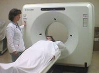
August 5, 2011 – New software aimed at making computed tomography (CT) safer for patients estimates the radiation risk based on age, gender and size rather than using the current “one size fits all” approach. The software program, tested on more than 6,500 scans, was introduced at the 2011 Joint Meeting of the American Association of Physicists in Medicine (AAPM) and Canadian Organization of Medical Physicists (COMP).
Physicians frequently prescribe CT imaging to help diagnose and treat diseases and injuries. But CT uses ionizing radiation, which can result in side effects, including an increased risk of developing cancer later in life. Because CT scans are an important medical tool, scientists are continually trying to balance the minimum radiation dose that provides clear images without exposing the patient to more radiation than necessary.
Currently, the radiation dose a patient receives is based on the radiation output indicated by the CT machine – a “one size fits all” calculation. In reality, the amount of radiation absorbed by a patient varies according to age, gender and size, as well as the part of the body that is being imaged. Radiation is riskier for patients who are younger (particularly children), smaller and female. Although the risks of radiation are greater for some patients, there has been no way to measure that risk accurately until now.
“This is the first dose monitoring program that factors age, gender and patient size, as well as the organ being imaged, into the risk calculation,” said Ehsan Samei, Ph.D., lead author of the study and chief physicist at Duke University Medical Center, Durham, N.C. “This helps us to estimate each patient’s dose level and potentially a more reliable level of risk for each patient. Having a more patient-specific estimate of risk can help to make CT safer.”
In the study, researchers used the program to analyze more than 6,500 CT scans of the chest, abdomen/pelvis and head performed during a five-week period at two hospitals. The new software factors in the radiation output noted on the CT machine, as well as the patient’s age, gender, size and organ being imaged to calculate the effective dose and an index of risk specific to the patient
Radiation risk is greater for younger patients, because they have more years ahead of them during which long-term effects could eventually develop. Because children are growing rapidly, their cells are more vulnerable to radiation damage. Conversely, the older the patient, the less damaging the radiation is. Although scientists don’t know exactly why, females are almost twice as susceptible to damage from radiation as males, unrelated to size and even the organ being imaged. And radiation absorption to certain areas of the body – such as the abdomen – are higher than to others, such as the head.
“This system quantifies the risks encountered by individual patients,” said Olav Christianson, co-author of the study and a medical physicist at Duke. “It is designed based on these very patient-specific factors to guide doctors in determining the most appropriate radiation dose patients should receive during a CT exam.”
For more information: www.aapm.org, www.medphys.ca


 January 05, 2026


