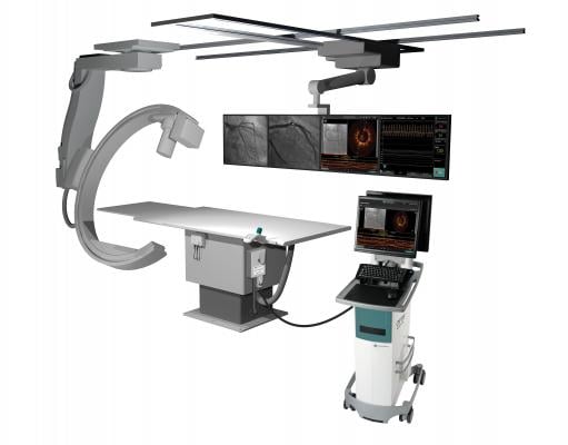
February 8, 2016 — St. Jude Medical Inc. announced the launch of the company’s Optis Mobile System in Japan and Europe. The diagnostic system is designed to couple state-of-the-art optical coherence tomography (OCT) and angiography co-registration with fractional flow reserve (FFR) technology into one portable system for hospitals with multiple catheterization labs.
Driven by a global increase in vascular disease and enabled by advances in technology, the catheterization lab allows physicians to use minimally invasive percutaneous coronary intervention (PCI) procedures that give patients an alternative to open heart surgery; the goal is to improve patient outcomes, shorten hospital stays and reduce hospital costs. The latest Optis System offers physicians an efficient way to optimize PCI procedures for the treatment of vascular disease by combining diagnostic tools designed to improve patient outcomes into one portable device.
Utilizing a combination of technology previously available only in the St. Jude Medical Optis Integrated System, the Optis Mobile System is designed to help physicians make improved stenting decisions based on high-resolution and three-dimensional OCT views of coronary anatomy while simultaneously mapping their exact location via angiogram. The system also integrates St. Jude Medical PressureWire fractional flow reserve (FFR) measurement technology, which offers physicians detailed coronary hemodynamic (circulatory) information during PCI. Clinical data has shown the physiological measurements provided by FFR can improve outcomes and reduce healthcare costs over traditional diagnostic imaging tools.
“As the interventional cardiology landscape continues to expand, there is a real need for more portable intravascular imaging systems to ensure hospitals with multiple catheterization labs have the right technology available for physicians to make more informed treatment decisions during PCI,” said Nick West, M.D., Papworth Hospital, Cambridge, U.K. “The imaging advancements offered with the Optis Mobile System provide the same benefits of the Optis Integrated System, and allow physicians to clearly visualize complex cardiac anatomy and evaluate how to best proceed during PCI.”
Sometimes referred to as coronary angioplasty, PCI is a catheter-based procedure designed to open coronary blockages or plaque buildup and restore blood flow to the heart. Traditionally, physicians have relied on either angiography or intravascular ultrasound to guide PCI procedures. St. Jude Medical’s Ilumien System first combined OCT and FFR technology to enable a more detailed, physiological and anatomical analysis of blood flow blockages inside the coronary vessels.
St. Jude Medical’s OCT technology is an intravascular imaging tool that uses light to provide anatomical images of disease morphology and automated measurements. With OCT technology, physicians can visualize and measure important vessel characteristics that are otherwise not visible or difficult to assess with the older imaging technology. As a result, OCT can provide automated, highly accurate measurements that can help guide stent selection and deployment and assess stent placement to help ensure successful procedures. This can potentially minimize the need for repeat revascularization.
FFR is a physiological index used to determine the hemodynamic severity of narrowings (or lesions) in the coronary arteries, and is measured using St. Jude Medical PressureWire Aeris and PressureWire Certus systems. FFR specifically identifies which coronary narrowings are responsible for obstructing the flow of blood to a patient’s heart muscle (called ischemia), and helps guide the interventional cardiologist in determining which lesions warrant stenting, resulting in improved patient outcomes and reduced health care costs.
For more information: www.sjm.com


 October 24, 2025


