May 21, 2019 – Medical diagnostic artificial intelligence (AI) company MaxQ AI announced that Accipio Ax will begin ...
Cardiac Imaging
The cardiac imaging channel includes the modalities of computed tomography (CT), cardiac ultrasound (echocardiography), magnetic resonance imaging (MRI), nuclear imaging (PET and SPECT), and angiography.

 May 21, 2019
This is a sample of the 3-D printed hearts and coronary anatomy models created from patient CT scans to enable ...
May 21, 2019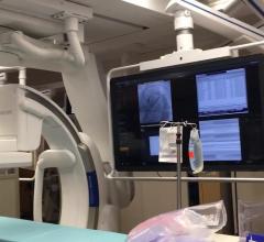
This is a walk through of the primary structural heart hybrid cath lab at Henry Ford Hospital in Detroit, Mich. It is ...
May 20, 2019Sponsored Content

Feature
SPONSORED CONTENT — Studycast is a comprehensive imaging workflow system that allows healthcare professionals to work ...
June 17, 2025
A demonstration of how to calculate the neo-left ventricular outflow tract (neo-LVOT) on CT imaging for a transcatheter ...
May 20, 2019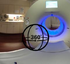
This is a 360 degree photo of a Siemens Somatom Force 64-slice, dual-source computed tomography (CT) system installed at ...
May 20, 2019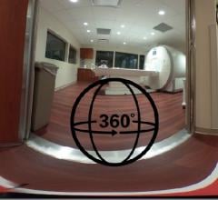
This is a dedicated cardiac Siemens 1.5T MRI system installed at the Baylor Scott White Heart Hospital in Dallas. The ...
May 17, 2019Sponsored Content
Feature | Cardiac Imaging
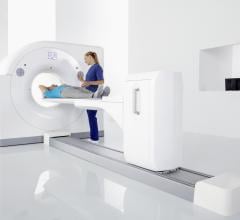
Cardiac positron emission tomography (PET) is growing in popularity among cardiologists because it provides the ability ...
March 05, 2024
May 17, 2019 ― Miami Cardiac & Vascular Institute announced the implementation of Philips’ Ingenia Ambition 1.5T MR, the ...
May 17, 2019
May 17, 2019 — Biopharmaceutical company CellPoint plans to begin patient recruitment for its Phase 2b cardiovascular ...
May 17, 2019
This is an example of how the heart's left atrial appendage (LAA) can be evaluated for thrombus and possible ...
May 16, 2019Sponsored Content

Blog | Enterprise Imaging
As medical advancements continue to push the boundaries of what is possible in the field of structural heart ...
August 10, 2023
This is an example of a carotid artery reporting module from Change Healthcare at 2018 Radiological Society of North ...
May 16, 2019
May 15, 2019 — Artificial intelligence (AI) solutions provider Aidoc has been granted U.S. Food and Drug Administration ...
May 15, 2019
May 13, 2019 — Imricor announced the signing of a commercial agreement with the Haga Hospital in The Hague, Netherlands ...
May 13, 2019Sponsored Content

Feature | Cardiac Imaging
Discover the key features of cardiovascular structured reporting that drive adoption, including automated data flow, EHR ...
November 07, 2022
The integration of artificial intelligence (AI) into medicine has by far been the hottest topic at nearly all medical ...
May 13, 2019
News | Radiopharmaceuticals and Tracers | Jeff Zagoudis, Associate Editor
May 10, 2019 — Shine Medical Technologies Inc. broke ground on their first medical isotope production facility in ...
May 10, 2019
May 9, 2019 — Osprey Medical announced the launch of DyeMINISH, a global patient registry to evaluate the ongoing safety ...
May 09, 2019