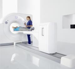
As hospital imaging departments look to replace aging nuclear scanners with updated technology, many are asking if ...
Cardiac Imaging
The cardiac imaging channel includes the modalities of computed tomography (CT), cardiac ultrasound (echocardiography), magnetic resonance imaging (MRI), nuclear imaging (PET and SPECT), and angiography.

February 14, 2020 – The American Society of Nuclear Cardiology (ASNC) and the American Society of Echocardiography (ASE) ...
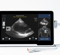
February 13, 2020 — The U.S. Food and Drug Administration (FDA) cleared software to assist medical professionals in the ...
SPONSORED CONTENT — Studycast is a comprehensive imaging workflow system that allows healthcare professionals to work ...
February 12. 2020 — Philips announced a new randomized controlled trial to assess patient outcomes after receiving a ...
February 12, 2020 — The University of Wisconsin (UW) Health’s University Hospital in Madison, Wis., recently became the ...

February 7, 2020 – At the 2019 Radiological Society of North America (RSNA) meeting in December, there was a record ...
Cardiac positron emission tomography (PET) is growing in popularity among cardiologists because it provides the ability ...
January 29, 2020 – The presence of a blood clot on the wall of the aorta in people with abdominal aortic aneurysms (AAA) ...
The key question I am always asked at cardiology conferences is what are the trends and interesting new technologies I ...

Cardiology was already heavily data driven, where clinical practice is driven by clinical study data, but mining a ...
As medical advancements continue to push the boundaries of what is possible in the field of structural heart ...
DAIC/ITN Editor Dave Fornell takes a tour of some of the most innovative new medical imaging technologies displayed on ...
January 9, 2020 — Maulik Majmudar, M.D., chief medical officer at Amazon will be the keynote speaker at the upcoming ...
January 7, 2020 — The American Society of Echocardiography (ASE) has updated its guidelines for using echocardiography ...
Discover the key features of cardiovascular structured reporting that drive adoption, including automated data flow, EHR ...
Karen Ordovas, M.D., MAS, professor of radiology and cardiology at the University of California San Francisco (UCFS) ...
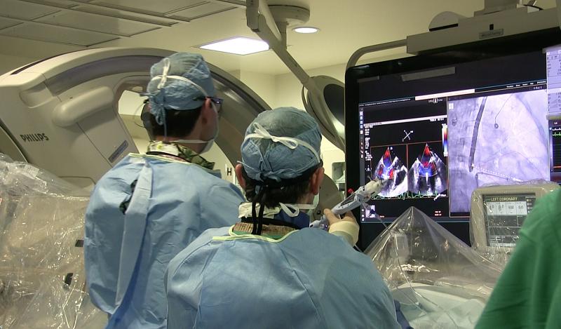
December 30, 2019 — Acoustoelectric cardiac imaging, a new, noninvasive cardiac imaging technology developed at the ...

 February 19, 2020

