December 3, 2021 — Here is the list of the most popular content on the Diagnostic and Interventional Cardiology (DAIC) m ...
Cardiac Imaging
The cardiac imaging channel includes the modalities of computed tomography (CT), cardiac ultrasound (echocardiography), magnetic resonance imaging (MRI), nuclear imaging (PET and SPECT), and angiography.
December 2, 2021 — Artificial intelligence (AI) vendor DiA Imaging Analysis was featured in a recent study presented by ...
December 1, 2021 — A small but significant percentage of college athletes with COVID-19 develop myocarditis, a ...
SPONSORED CONTENT — Studycast is a comprehensive imaging workflow system that allows healthcare professionals to work ...
Examples of TrueView and GlassView 3D cardiac ultrasound visualization and artificial intelligence (AI) assisted ...
November 22, 2021 — The Minneapolis Heart Institute Foundation (MHIF) announced the publication of research showing ...
Dr. Simon Dixon, MBChB, chair of the Department of Cardiovascular Medicine at Beaumont Hospital Royal Oak, the Dorothy ...
Cardiac positron emission tomography (PET) is growing in popularity among cardiologists because it provides the ability ...
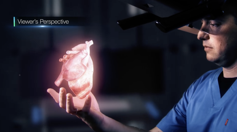
November 15, 2021 — RealView Imaging Ltd. recently received FDA 510(k) clearance for its Holoscope-i holographic system ...
November 10, 2021 — Philips Healthcare announced North American availability of new innovations in its portfolio of ...
November 10, 2021 — Philips, a global leader in health technology, announced that it has signed an agreement to acquire ...
As medical advancements continue to push the boundaries of what is possible in the field of structural heart ...
November 9, 2021 — Caption Health and Ultromics, developers of artificial intelligence (AI) to improve cardiac ...
November 5, 2021 – An FDA-approved device used during cardiac cath lab procedures cut radiation exposure for ...
November 1, 2021 — According to ARRS’ American Journal of Roentgenology (AJR), radiologists need to be cognizant of the ...
Discover the key features of cardiovascular structured reporting that drive adoption, including automated data flow, EHR ...
November 1, 2021 — Here is the list of the most popular content on the Diagnostic and Interventional Cardiology (DAIC) ...

October 29, 2021 — A new guideline for the evaluation and diagnosis of chest pain was released this week that provides ...
October 20, 2021 — HeartFlow Inc. announced the commencement of the REVEALPLAQUE (A pRospEctiVe, multicEnter study to ...

 December 03, 2021


