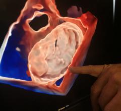
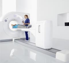
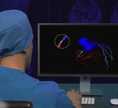
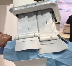
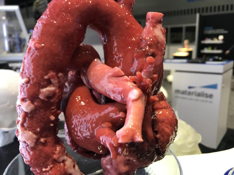
The gallery includes photos of medical imaging technologies from across the expo floor at the Radiological Society of ...
Cardiac Imaging
The cardiac imaging channel includes the modalities of computed tomography (CT), cardiac ultrasound (echocardiography), magnetic resonance imaging (MRI), nuclear imaging (PET and SPECT), and angiography.

Here are a few of the key takeaways on the new technologies that will impact cardiovascular and interventional medicine ...
Orlando Simonetti, Ph.D., professor, cardiovascular medicine, worked with Siemens to help develop a new, lower-field mag ...
SPONSORED CONTENT — Studycast is a comprehensive imaging workflow system that allows healthcare professionals to work ...
January 18, 2022 – Philips Healthcare announced physicians will now have access to advanced new 3D image guidance ...
January 17, 2022 – As the increasing number of structural heart interventions are assisted by real-time imaging guidance ...
Here are two examples of artificial intelligence (AI) driven pulmonary embolism (PE) response team apps featured by ...
Cardiac positron emission tomography (PET) is growing in popularity among cardiologists because it provides the ability ...
January 4, 2022 - Pie Medical Imaging (PMI), a global leader in cardiac imaging, part of the Esaote Group, recently ...
Wishing you a healthy and happy New Year from the DAIC team. Look for many exciting new changes to come in 2022!
December 23, 2021 — An interdisciplinary research team from the University of Göttingen and Hannover Medical School (MHH ...
As medical advancements continue to push the boundaries of what is possible in the field of structural heart ...
December 14, 2021 — RSIP Vision, a medical imaging company applying advanced artificial intelligence (AI) and computer ...
Jean Jeudy, M.D., professor of radiology and vice chair of informatics at the University of Maryland School of Medicine ...
December 13, 2021 - HeartFlow Inc., the leader in revolutionizing precision heart care, today announced it has submitted ...
Discover the key features of cardiovascular structured reporting that drive adoption, including automated data flow, EHR ...
The vendor Radiaction introduced a new type of scatter radiation protection shielding system that mounts to the ...
December 9, 2021 – Philips Healthcare expanded its cardiac ultrasound portfolio with new imaging tools and features to ...
Kate Hanneman, M.D., MPH FRCPC, director of cardiac imaging research JDMI, and the medical imaging site director at ...

 January 21, 2022


