April 20, 2022 – Researchers from the RIKEN Center for Advanced Intelligence Project (AIP) and colleagues have tested ...
Cardiac Imaging
The cardiac imaging channel includes the modalities of computed tomography (CT), cardiac ultrasound (echocardiography), magnetic resonance imaging (MRI), nuclear imaging (PET and SPECT), and angiography.

 April 20, 2022
April 13, 2022 — The Society of Nuclear Medicine and Molecular Imaging (SNMMI) and the American Society of Nuclear ...
April 13, 2022
April 12, 2022 — The American College of Cardiology has issued an expert consensus decision pathway for the evaluation ...
April 12, 2022Sponsored Content

Feature
SPONSORED CONTENT — Studycast is a comprehensive imaging workflow system that allows healthcare professionals to work ...
June 17, 2025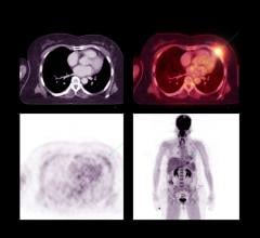
April 11, 2022 — Radiation to the heart during treatment for locally advanced lung cancer is associated with an ...
April 11, 2022
April 6, 2022 – According to ARRS’ American Journal of Roentgenology (AJR), the 3-T Dixon gradient-recalled echo (GRE) ...
April 06, 2022
April 6, 2022 – Myocarditis (inflammation of the heart muscle) is usually caused by viruses, such as in Covid-19 disease ...
April 06, 2022Sponsored Content
Feature | Cardiac Imaging
Cardiac positron emission tomography (PET) is growing in popularity among cardiologists because it provides the ability ...
March 05, 2024
April 2, 2022 — Aidoc, a leading provider of healthcare AI solutions, announced that it received FDA 510(k) clearance ...
April 02, 2022![Royal Philips, a global leader in health technology, today announced the launch of Ultrasound Workspace [OLK1] at the American College of Cardiology’s Annual Scientific Session & Expo (ACC 2022).](/sites/default/files/styles/content_feed_medium/public/philips-ultrasound-workspace-cardiology-diagnosis.jpeg?itok=VFUpaaT2)
April 2, 2022 — Royal Philips, a global leader in health technology, today announced the launch of Ultrasound Workspace ...
April 02, 2022
March 31, 2022 – Investigators from Cedars-Sinai have created an artificial intelligence-enabled tool that may make it ...
March 31, 2022Sponsored Content

Blog | Enterprise Imaging
As medical advancements continue to push the boundaries of what is possible in the field of structural heart ...
August 10, 2023
March 31, 2022 – DiA Imaging Analysis Ltd, a leading global provider of AI-powered ultrasound analysis software ...
March 31, 2022
March 29, 2022 – Cardiac computed tomography (CCT) is an accurate, noninvasive diagnostic test for obstructive coronary ...
March 29, 2022
March 29, 2022 – Impulse Dynamics, a company dedicated to innovative treatments for chronic heart failure, has ...
March 29, 2022Sponsored Content

Feature | Cardiac Imaging
Discover the key features of cardiovascular structured reporting that drive adoption, including automated data flow, EHR ...
November 07, 2022
March 11, 2022 – Is cardiac computed tomography (CT) as reliable as catheterization in patients with suspected coronary ...
March 11, 2022
March 9, 2022 — Fujifilm Sonosite, Inc., a world leader in point-of-care ultrasound (POCUS) solutions today announced it ...
March 09, 2022
February 21, 2022 – Acute coronary syndromes (such as heart attacks) and strokes are a leading cause of morbidity and ...
February 21, 2022