
July 13, 2022 — The Society of Cardiovascular Computed Tomography (SCCT) has released an update on the device ...
Cardiac Imaging
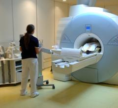
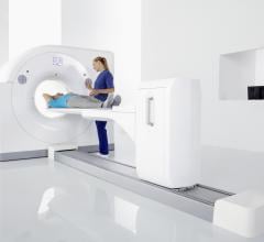
The cardiac imaging channel includes the modalities of computed tomography (CT), cardiac ultrasound (echocardiography), magnetic resonance imaging (MRI), nuclear imaging (PET and SPECT), and angiography.
July 11, 2022 – The American Society of Nuclear Cardiology (ASNC) and the Society of Nuclear Medicine and Molecular ...
 July 11, 2022
July 8, 2022 — The Society of Cardiovascular Computed Tomography (SCCT) has released a new expert consensus document on ...
July 08, 2022Sponsored Content

Feature
SPONSORED CONTENT — Studycast is a comprehensive imaging workflow system that allows healthcare professionals to work ...
June 17, 2025
Sponsored Content | Videos | Cardiovascular Ultrasound
Automated features on the Philips EPIQ CVx cardiology ultrasound system are helping to bring consistency and speed to ...
July 07, 2022
June 30, 2022 — Clarius Mobile Health, a leading provider of high-definition wireless ultrasound systems, and ImaCor Inc ...
June 30, 2022
June 29, 2022 — A spiral wave of electrical activity in the heart can cause catastrophic consequences. One spiral wave ...
June 29, 2022Sponsored Content
Feature | Cardiac Imaging
Cardiac positron emission tomography (PET) is growing in popularity among cardiologists because it provides the ability ...
March 05, 2024
June 27, 2022 — Three articles and an accompanying editorial provide information on the effects of Long COVID in the Deu ...
June 28, 2022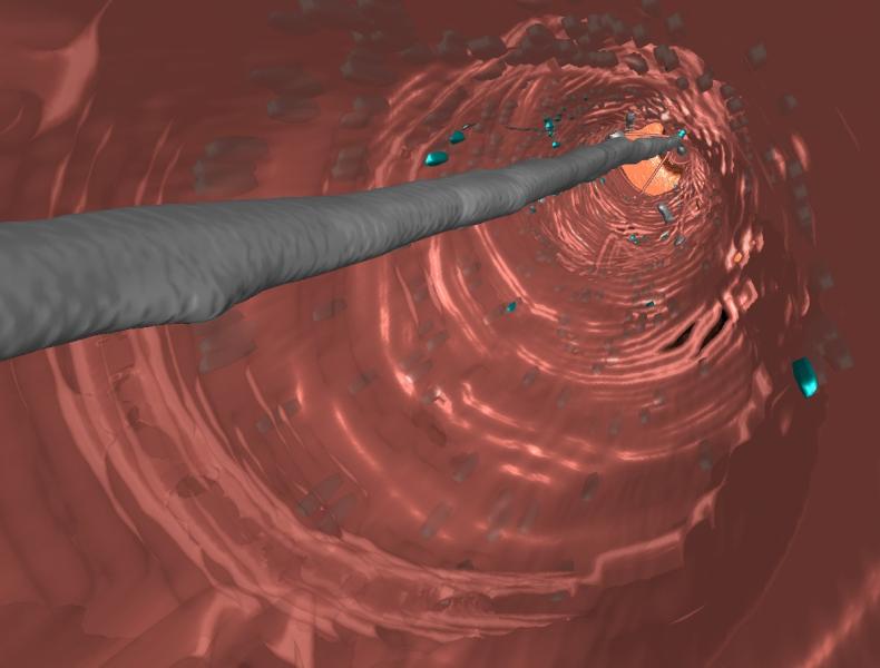
Feature | Cardiac Imaging | By Ronny Shalev, Ph.D.
Artificial intelligence’s (AI) applicability in cardiac imaging is rapidly growing and was a major topic of discussion ...
June 28, 2022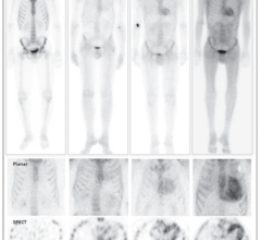
June 24, 2022 — Shortages of pyrophosphate (PYP), the radiopharmaceutical most commonly used in the U.S. for noninvasive ...
June 24, 2022Sponsored Content

Blog | Enterprise Imaging
As medical advancements continue to push the boundaries of what is possible in the field of structural heart ...
August 10, 2023
June 24, 2022 — The Radiological Society of North America (RSNA) reports that Coronary Artery Calcium (CAC) scoring with ...
June 24, 2022
June 23, 2022 — New research from the Smidt Heart Institute at Cedars-Sinai shows for the first time that the path ...
June 23, 2022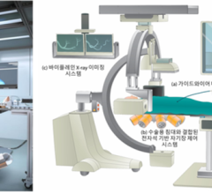
June 17, 2022 — A multidisciplinary team of robotics and electronic systems engineers working with cardiologists and ...
June 17, 2022Sponsored Content

Feature | Cardiac Imaging
Discover the key features of cardiovascular structured reporting that drive adoption, including automated data flow, EHR ...
November 07, 2022
June 16, 2022 — Xoran Technologies has recently received a patent for a modular computed tomography (CT) system assembly ...
June 16, 2022
June 15, 2022 — Cardiawave SA, a deeptech medical device manufacturer that has developed Valvosoft, a revolutionary non ...
June 15, 2022
June 15, 2022 — Seven finalists have been selected for the Best Abstract Award in cardiovascular computed tomography ...
June 15, 2022