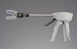
June 28, 2013 — Maquet Cardiovascular LLC announced it has acquired LAAx Inc., a privately held company that has developed a unique mechanical occlusion device called the TigerPaw System II. When implanted, TigerPaw II safely and effectively occludes the left atrial appendage (LAA) and conforms to its shape and thickness, minimizing the risk of tissue damage and accidental bleeding associated with other common closing methods used during open chest procedures. The product has been cleared by regulatory bodies in the United States and European Union and is commercially available in those regions.
"We believe TigerPaw II will be a best-in-class device for reliable ligation of the LAA, potentially mitigating post-operative stroke and its associated costs in patients with atrial fibrillation undergoing cardiac surgery," said Peter Hinchliffe, president and CEO for the cardiac surgery business unit of Maquet Cardiovascular. "Atrial fibrillation affects millions of people worldwide, and the Centers for Disease Control and Prevention expect that number to climb significantly in the years to come. We believe TigerPaw II will play an important role for our customers as they treat more and more patients with atrial fibrillation."
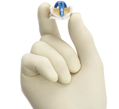
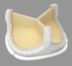
TigerPaw II is an implantable LAA occlusion device featuring an exceptionally soft silicone fastener that conforms to the appendage anatomy and tissue thickness, spreading the pressure equally. With its simple and reliable application, TigerPaw II is the only device currently available that has been proven 100-percent occlusive of the LAA during cardiac surgery, due to the pliable silicone.
"We are continuously evaluating and pursuing technologies that strengthen our offering to customers, and we believe this acquisition allows us to round out our cardiac surgery portfolio and provides us with an entry point into a growing market," said Raoul Quintero, president and CEO of Maquet Medical Systems USA. "TigerPaw II is our first product in what we hope will become a portfolio of products that helps address the needs of this important patient population."
Subsequent to the U.S. and E.U. launches, TigerPaw II will also become commercially available in other countries around the world following their local regulatory approvals.
The TigerPaw System II consists of a single-use delivery tool and an implantable fastener. The delivery tool has two triggers, one for positioning and placement of the fastener, and the second for release of the fastener, allowing delivery tool removal. The jaws uniquely include a 15-degree angle to allow the surgeon to gain easy and effective placement of the fastener. In a clinical trial, the placement of the fastener — hand in to hand off — was 60 seconds or less, with the average being 27 seconds. The delivery tool also allows for unlimited placement trials before actually having to apply the fastener.
The fastener is designed around the concept of interrupted mattress sutures, which are commonly used in friable, dynamic/compliant tissue environments. The fastener consists of a series of evenly spaced individual connectors embedded within a soft, compliant elastomeric housing. The compliant housing has three clinical benefits:
- Seals the puncture site of the needle barb, which penetrates both walls of the LAA ostium, thus eliminating leaking of blood through those sites;
- Allows for optimized conformance to dramatic variation of wall thickness, resulting in optimal occlusion performance; additionally, the housing material continues to adjust to tissue thickness variation as the affected site remodels and heals; and
- Infarcts the LAA, potentially eliminating atrial fibrillation triggers from originating within the LAA.
The TigerPaw System II is indicated for the occlusion of the LAA under direct visualization, in conjunction with other open cardiac surgical procedures. Direct visualization, in this context, requires that the surgeon is able to see the heart directly, without assistance from a camera, endoscope, etc., or any other viewing technology. This includes procedures performed by sternotomy (full or partial) as well as thoracotomy (single or multiple).
For more information: www.maquet.com


 January 15, 2026
