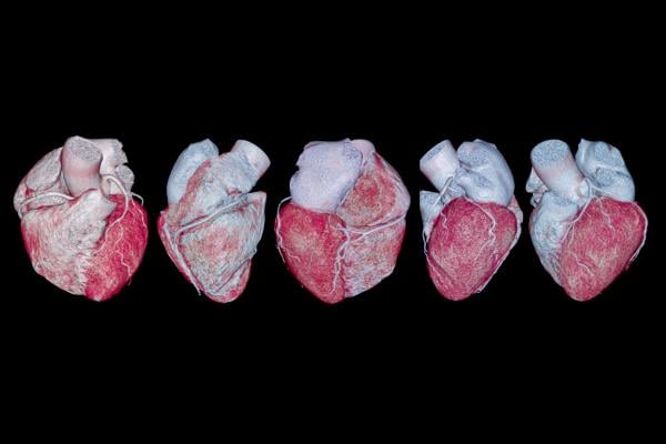
Cardiac CT scans, recommended by the American College of Cardiology (ACC) and the American Heart Association (AHA) as the primary testing strategy for patients with acute chest pain, are necessary for evaluating cases and determining treatment plans. Despite its importance, a substantial number of U.S. patients who require a cardiac CT miss out on receiving one due to limited access.
Cardiac CT isn't just a diagnostic tool — it's an essential component of cardiovascular health. Employing in-house cardiac imaging equips clinicians with enhanced insights into heart structures, allowing for more effective diagnosis and treatment, all while improving efficiency and the patient experience.
The traditional approach, relying on external referrals and involving multiple healthcare facilities, has several drawbacks. External referrals often result in prolonged patient wait times and delayed treatment. For providers, referring patients externally for imaging not only burdens patients but also results in missed revenue opportunities. Embracing the in-house model streamlines the entire process, providing a more seamless and personalized patient experience while offering healthcare providers a more lucrative opportunity.
The evolution of cardiac imaging solutions
In the early 2000s, 64-slice scanners marked a milestone in cardiac CT imaging. Over the years, subsequent scanners have introduced advancements, such as faster speeds, increased volume coverage, and enhanced capabilities. Modern scanners and techniques substantially reduce radiation and contrast agent doses.
Cardiovascular CT's non-invasive nature allows patients to avoid being admitted to a hospital for imaging, which makes it more convenient and patient-friendly. Invasive catheterizations, commonly employed, may fall short in identifying obstructions that prior CT imaging of the coronaries can catch.
Committed to advancing patient care, I introduced cardiac CT in my private practice, becoming the first private cardiologist in the region to do so. My goal was to inspire concrete behavioral changes and enhance patient adherence to treatment. Presenting patients with a tangible visualization of their current arterial condition, alongside a comparative analysis of others, proved more impactful than merely informing them about their conditions.
Improved patient safety with reduced radiation and contrast load
Enhancing patient safety is a pivotal aspect of advanced cardiac imaging technology. The substantial reduction in radiation exposure and contrast load is a leap forward in safety standards. By employing advanced scanning technology, healthcare providers can ensure patients undergo diagnostic procedures with minimized risks.
Compared to other scanners, Arineta’s SpotLight scanner offers a reduction in radiation exposure. Traditional scanners typically expose patients to considerable radiation amounts, ranging from 15 to 30 millisieverts. However, I usually scan patients with less than 2-3 millisieverts using the SpotLight. For context, living on Earth exposes us to around five to six millisieverts of radiation each year.
With the same scanner, we also reduce the contrast load from the usual 80 to 100 mLs down to 45 to 55 mLs. As a result, we’re cutting the contrast load in half, lowering the risk of kidney problems and other contrast-related issues for patients. Lowering both contrast and radiation levels enables more frequent scanning for patients with moderate or stage two and three coronary artery disease.
Advanced scanners provide detailed insights into plaque progression and stabilization. As a result, we can provide more proactive patient care. For instance, if a patient has a blockage in the 60% to 70% range, is asymptomatic, and stress testing is negative, and there's no indication for a catheter, we can employ aggressive medical therapy and then re-scan the patient in a year. By doing so, we can monitor plaque progression or stabilization.
Direct examination of the arteries through advanced imaging provides a thorough assessment of cardiovascular conditions, leading to more precise diagnoses and personalized treatment plans. By shifting away from indirect surrogate markers, diagnostic accuracy is amplified, and uncertainty in patient care is minimized. A clearer view of the arterial system enables healthcare professionals to identify and address issues more effectively, contributing to improved patient outcomes.
Referral process and challenges
When patients need cardiac imaging but lack access to an in-house facility, they are often referred to hospitals or outpatient centers. With external referrals, patients lack personalized experiences. They navigate through a complex system where they encounter multiple healthcare professionals at various stages of the imaging process, leading to a maze of appointments.
Patients opting for cardiac imaging in a hospital setting, while essential for many, often experience prolonged wait times for appointments. The intricacies of scheduling within large healthcare facilities and the high demand for imaging services contribute to delays in securing a slot for necessary diagnostic procedures. When there isn't a specific and reliable point of contact, misunderstandings and delays can occur.
In contrast, opting for in-house cardiac imaging provides a more efficient and streamlined experience, resulting in reduced wait times and a more personalized process. With dedicated imaging facilities within a specialized practice, patients can easily secure timely appointments, enabling prompt diagnosis and treatment planning.
Benefits of in-house cardiac imaging
In-house cardiac imaging offers a centralized and streamlined diagnostic process, reducing wait times and facilitating quicker assessments for timely treatment decisions. The absence of external referrals means patients receive comprehensive care in a familiar setting, leading to increased satisfaction and building trust between the patient and healthcare provider.
Having an in-house facility not only enhances efficiency but also allows clinicians to deliver a more comprehensive and tailored approach to patient care. Timely imaging enables the early identification of cardiac conditions, paving the way for proactive intervention and ultimately resulting in improved patient outcomes.
For instance, a 42-year-old patient with a family history of heart issues received reassurance from various cardiologists over the years, as his normal stress test results suggested a low risk. However, when we conducted an in-office coronary CT angiogram, it revealed blockages in all three blood vessels, despite prior assurance of normal arteries.
Our unexpected discovery revealed the patient had stage three coronary artery disease, which indicates a greater than 15% chance of a heart attack or death within 10 years. The significant difference between the results of the stress tests and the CT angiogram highlights the importance of direct testing in accurately assessing cardiovascular risk.
Financial impact and overcoming barriers to adoption
Beyond the evident benefits for patients, the integration of in-house advanced imaging has proven to be an effective financial decision, leading to a substantial 40% increase in revenue for my practice. The presence of an in-house CT scanner not only elevates diagnostic capabilities but also contributes to the overall financial stability.
A key advantage is the practice's capacity to attract patients from different areas who value the convenience and efficiency of in-house imaging services. An on-site CT scanner minimizes reliance on external referrals, ensuring a smoother experience for patients and establishing the practice as a comprehensive destination for cardiac care.
For providers contemplating the adoption of in-house cardiac imaging, having an in-house reader enhances the efficiency and effectiveness of the imaging program. The presence of a dedicated professional on-site streamlines the interpretation process, reducing turnaround times, and improving overall diagnostic accuracy.
Another important factor is understanding where the patient volume comes from. Before introducing in-house imaging, it's necessary to thoroughly grasp patient demographics, referral patterns, and potential imaging needs. Armed with this information, providers can customize their program to align with the specific demands of the community and referring physicians, ensuring a more focused and successful implementation.
Future directions and technological advancements
In less than 10 years, I transitioned from a small 1,100-square-foot office to a larger 3,500-square-foot space, now serving 50 to 100 patients daily. Initially conducting four to 10 cardiac CT scans weekly, our practice evolved considerably through community outreach, physician education, and referral base development. We've successfully established a streamlined in-house cardiac imaging practice, performing 60 to 100 CT scans weekly.
Cardiology is moving toward direct testing, and advanced CT scanners are an important step in that direction. Modern scanners not only produce high-resolution images for precise diagnoses but also reduce radiation and contrast loads, aligning with the growing emphasis on patient safety and comfort.
Advanced scanners are a major advancement in cardiac imaging, allowing more frequent assessments to track patients' progress. The proactive use of regular imaging provides a dynamic understanding of cardiovascular health, allowing for timely interventions and personalized care plans, departing from the traditional sporadic imaging approach.
The future of cardiology will rely on technological advancements to enhance diagnostic precision and improve patient outcomes. The incorporation of state-of-the-art CT scanners and a move toward direct testing indicate a shift in cardiovascular care, where innovation and patient-focused approaches come together for more effective healthcare delivery. Our use of advanced technology reflects the evolving standards in cardiology.

Alberto Morales, MD, is a cardiologist and owner of South Tampa Cardiology and Advanced Imaging Center in Tampa, Fla. Committed to providing South Tampa with the highest quality of cardiology care, Dr. Morales invested in building one of the most advanced, free-standing cardiology and imaging centers in the region. By combining his seasoned cardiology team and the latest state-of-the-art imaging technology, all under one roof, patients can consult with the doctor and undergo life-saving imaging all in one convenient location.
Related Cardiac CT content:
The Evolving Computed Tomography Market
AI Breakthrough Award Presented to Cleerly CCTA Digital Care Platform
HeartFlow to Announce New Data at SCCT23
The Benefits of Coronary CTA in Pharmaceutical Clinical Trials
Photon-counting CT Noninvasively Detects Heart Disease in High-risk Patients
Rising Demand for Cardiac CT Positions Market for Major Growth


 November 14, 2025


