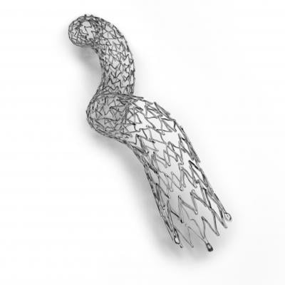
October 5, 2018 — Veryan Medical Ltd has received Premarket Approval (PMA) for the BioMimics 3D Vascular Stent System from the U.S. Food & Drug Administration (FDA). The device is approved for the treatment of symptomatic de novo or restenotic lesions in the native superficial femoral artery and/or proximal popliteal artery.
The BioMimics 3D stent has a three-dimensional helical shape, designed to impart natural curvature to the diseased femoropopliteal artery, to promote swirling flow and elevate wall shear, which has a protective effect on the endothelium.1,2, 3 The helical shape of the stent is also designed to facilitate shortening of the stented segment during knee flexion and mitigate the risk of stented segment compression causing localized strains that in a straight stent may lead to stent fracture and chronic vascular injury.5, 6
Key components of the PMA application were the 12-month interim safety and effectiveness results from the company’s MIMICS-2 clinical study. The study was conducted under an FDA-approved Investigational Device Exemption (IDE) in patients with peripheral arterial disease (PAD) undergoing endovascular intervention in the femoropopliteal artery.
The company said BioMimics 3D represents an innovative approach to the requirement for durable support for the arterial lumen after intervention. The helical centerline stent is designed to not only promote swirling blood flow but also to accommodate the complex biomechanical challenge associated with stenting this anatomically mobile artery.
The MIMICS-2 study enrolled 271 subjects across 43 investigational sites in the U.S., Japan and Germany. The principal investigators are Timothy M. Sullivan, M.D., Minneapolis; Masato Nakamura, M.D., Ph.D., Tokyo, Japan; and Thomas Zeller, M.D., Bad Krozingen, Germany.
Both primary endpoints in the MIMICS-2 study, safety and effectiveness, were met. Freedom from major adverse events at 30 days was 99.6 percent (268/269) and Kaplan-Meier (KM) estimates of freedom from loss of primary patency and clinically-driven target lesion revascularization (CDTLR), were 83 percent and 88 percent, respectively, at 12 months; no stent fractures were detected in core laboratory imaging review.4
For more information: www.veryanmed.com
References
- Zeller T. - Oral Presentation VIVA 2014
- Malek, JAMA, 282, p 2035-2042, 1999
- Zeller T. et al; Circ Cardiovasc lnterv. 2016;9:e002930. DOI: 10.1161 2
- Data on file at Veryan Medical
- BH Smouse et al, Endovasc. Today, vol 4, no. 6, pp. 60-66, 2005
- Scheinert D et al, J Am Coll Cardiel 2005;45:312-5 doi:10.1016/j.jacc.2004.11.026


 November 14, 2025


