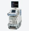
April 8, 2009 - The Helena Cardiology Clinic of Helena, MT has installed two Toshiba Aplio Artida ultrasound systems with 3D Wall Motion Tracking capabilities, allowing sonographers and physicians to more quickly and accurately identify wall motion defects and the timing of cardiac events.
The feature is designed to greatly improve the detection of wall motion abnormalities in many cardiac disease states and cardiac resynchronization Therapy (CRT) and helps physicians optimize pacemaker settings.
“This advancement in imaging will enable me to better manage the care of my cardiac patients,” said Richard Paustian, M.D., owner of the Helena Cardiology Clinic. “In addition, these added clinical benefits enable us to expand our service offerings and provide a wider range of patients with advanced treatment.”
Using Artida’s real-time, multi-planar reformatting capabilities, physicians can reportedly assess global and regional LV function, including volumetric LV ejection fraction. Arbitrary views of the heart, not available in 2D imaging, are also obtained to help with surgical planning. The 3D Wall Motion Tracking features allow the user to obtain angle-independent, global and regional information about myocardial contraction. It is hoped these features will enable acquisition of additional data that could be of value in echo-guided CRT and in stress echocardiography.
For more information: www.medical.toshiba.com.


 January 28, 2026


