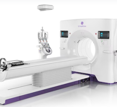
The University of Virginia Health System uses a Siemens MAGNETOM Avanto 1.5T MRI system.
"Potential” can be a loaded word, especially in healthcare. Many clinicians have tagged coronary MR angiography (MRA) with the “potential” label. Despite the scan’s reported safety advantages over coronary CT angiography (CTA), as well as its superior physiologic information, coronary MRA has not gained the widespread acceptance of coronary CTA.
Yet, many clinicians are hopeful the scan will gain momentum in the coming years as new techniques and new hardware/software progress.
What is the Potential?
Many cardiologists and radiologists are excited about the potential for coronary MRA to evaluate plaque. MR has been able to see and evaluate plaque for years in the carotid, aortic and peripheral arteries, according to Warren Manning, M.D., professor of radiology at Harvard Medical School, member of the cardiovascular disease department at Beth Israel Deaconess Medical Center in Boston. However, this potential has not led to significant clinical applications for coronary MRA for a large patient population.
“Plaque in coronary arteries is far more complicated in the MR,” Dr. Manning said. “The coronary wall is even smaller than the coronary lumen, so the burdens are more strict with motion compensation. There are techniques using gadolinium looking at inflammation in the wall of the arteries.
“CT looks at the anatomy. Is there a calcified plaque or a soft plaque? MR is looking for any plaque or if there is inflammation in the wall. We think inflammation probably means someone is at an increased risk for plaque rupture, but at the present time it’s just a hypothesis. We don’t really know for sure that MRI can identify the sites where plaque will rupture. All of this for [MR] coronary plaque imaging is really in the research arena at the present time.”
Safety, Imaging Advantages
Currently, coronary MRA’s primary advantage over CTA is that it doesn’t use radiation or iodinated contrast material.
“One of the issues that often comes up is if patients are going to need repeated studies,” Dr. Manning said. “Repeated studies in the CT world mean repeated exposure to radiation. There are no such issues with MR.”
Many doctors, as well as parents, are hesitant to expose children to radiation from a CT scan. For this reason, coronary MRA is preferred for younger patients being imaged for anomalous coronary artery disease, according to an American Heart Association Scientific Statement released in July.
“If one isn’t trying to discriminate ischemic versus non-ischemic cardiomyopathies, then I think both MR and CT work, and we’d say that MR may be preferred in that group as well,” Dr. Manning said.
Coronary MRA doesn’t typically use contrast, while CTA uses iodine-based contrast materials that can be problematic for patients with renal insufficiencies.
The scan also holds advantages over CTA in that it doesn’t require a slow heartbeat and it images calcium better. Heavily calcified arteries are a burden for CTA. However, in part because the MR allows for free breathing, a coronary MRA has an acquisition time of 5-10 minutes, as opposed to less than 20 seconds for a CTA.
Spatial Resolution vs. CTA
The major drawback to coronary MRA is the inferior spatial resolution, according to Christopher M. Kramer, M.D., professor of radiology and medicine, director of cardiovascular imaging, University of Virginia Health System.
“Because CT has higher spatial resolution and has performed better for stenosis detection, CTA is preferable,” Dr. Kramer said. “For many other cardiac applications, MR is preferable. In 2008, for assessment of coronary artery disease in a middle- aged or older patient, CT is probably preferable.”
Dr. Manning believes that although CTA has better spatial resolution, a coronary MRI scan is still very readable.
“If you have experience [in reading both scans], then I think the interpretation is similar,” Dr. Manning said. “I think there are more 64-slice CT scanners in existence than there are MR sites that have experience in coronary MRI. People need to consider their local expertise like every other imaging or procedural modality.”
Coronary MRI has improved in recent years, according to Dr. Kramer, who has used Siemens’ MAGNETOM Avanto 1.5T MRI system since 2004.
“The major improvements have been the use of 3D coronary imaging,” Dr. Kramer said. “This is a 3D whole-heart approach where a navigator is used to track diaphragmatic motion and capture the 3D image of the heart over many heart beats. It’s respiratory-gated using navigator echoes. The improvement in the navigators has also helped image quality.”
Dr. Manning, who uses a Philips 1.5T MRI system, also noted scanners have moved from 16 to 32 channel coils, which helps with signal-to-noise ratios.
New Technology, Techniques
There is no shortage of ways in which clinicians are looking to improve spatial resolution to help coronary MRA reach its potential.
“Going to 3 Tesla has the potential to improve image quality and spatial resolution for certain applications,” Dr. Kramer said. “Coronary imaging is one of them. There’s certainly more data coming out now with whole-heart coronary angiography MR at 3T. I think the performance will be better than at 1.5T, but still not to CTA spatial resolution.”
Dr. Manning noted than MR can yield better results without having a hardware upgrade.
“MR advances are very much software-based,” Dr. Manning said. “New algorithms to do something. New sequences. But, you’re still using the same 1.5T or 3T scanner. That’s another advantage of MR.”
Other solutions to the spatial resolution problem include using a gadolinium-based contrast for the coronaries, using an intravascular MRI or developing the scan on higher field MR system.
“There is some work on intravascular MRI, but that’s invasive and not useful for a general patient population screening,” Dr. Kramer said. “There are 7T scanners scattered around the world, but there are a lot of issues to be tackled when you go to that high of a field.
“There is a potential to use MR contrast in imaging the coronary arteries,” he said. “It’s a little difficult because it’s a long acquisition, so the timing of contrast infusion relative to the acquisition of the images is tricky, but it may enhance coronary imaging.”


 February 02, 2026


