October 9, 2007 - From a snippet of a patient’s skin, researchers have grown blood vessels in a laboratory and then implanted them to restore blood flow around the patient’s damaged arteries and veins.
It is the first time blood vessels created entirely from a patient’s own tissues have been used for this purpose, the researchers report in the current issue of The New England Journal of Medicine.
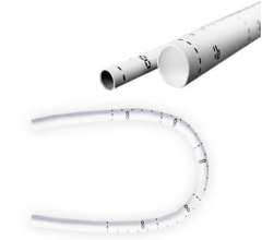
Cytograft Tissue Engineering of Novato, Calif., made the vessels, in a process that takes six to nine months. Because they are derived from patients’ own cells, they eliminate the need for antirejection drugs. And because they are devoid of any synthetic materials or a scaffolding, they avoid complications from inflammatory reactions.
Doctors in Argentina have performed the first human tests of the vessels on six patients, the team reported. Two additional implants have been performed since the report was submitted, said Dr. Todd N. McAllister of Cytograft.
The longest follow-up among these patients has been for 13 months. Cytograft says the vessels hold promise for patients with damaged blood vessels from diabetes, arteriosclerosis, birth defects and other problems. But monitoring for a much longer period will be needed before those uses could become standard, the team said.
“This technique has a big potential in the vascular surgical field,” said Dr. Toshiharu Shinoka, who directs pediatric cardiovascular surgery at Yale and who plans to conduct studies with Cytograft on the new vessel. He called the technique an advance over one he used in operations on children in Japan, in which vessels were grown from cells on a scaffold that then degraded and was absorbed into the body.
Doctors not connected with the company agreed on the importance of the new technique. “A potential benefit may be for infants and children with congenital heart defects,” said Dr. Deepak Srivastava, director of the Gladstone Institute of Cardiovascular Disease at the University of California, San Francisco. Unlike grafts from cadavers, he added, “the Cytograft vessels should be able to grow as the child does.”
The Cytograft studies were done at a leading cardiovascular center in Argentina, where medical costs are much lower than in the United States. The patients were all receiving chronic kidney dialysis, which cleanses wastes from the blood.
As is standard for such treatment, doctors had surgically cut an artery and a vein in the forearm and joined them in a link known as a shunt, which provides access for the repeated needle punctures needed to connect a patient to a dialysis machine. Such shunts can last up to 15 years. But when clots and infections develop in the shunts to reduce or stop blood flow, new ones must be created.
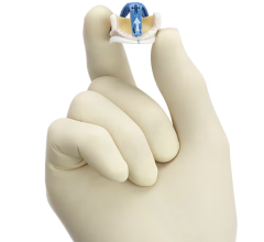
Dr. Sergio A. Garrido, a vascular surgeon in Buenos Aires, said he implanted the Cytograft vessels in the forearm or upper arm under general anesthesia, in a different area from the malfunctioning shunt. The procedure took 60 to 90 minutes. Through surgical gloves, the Cytograft vessel, 5 ½ to 11 ¾ inches long, felt a little more delicate than a regular vein, he said.
Volunteers were chosen for the study because of their need and because the shunts allowed an easy way to monitor the functioning of the new vessels. The doctors performed ultrasound tests three times a week and could repeatedly puncture the vessels for dialysis.
Vessels in the arm would be much easier to repair in an emergency than vessels implanted deeper in the body. In one potential use of the technique, the team hopes to use the vessels to save the toes and lower legs of people with poor blood circulation from arteries damaged by diabetes and arteriosclerosis. A second potential use is among people who need coronary bypasses. Cytograft is recruiting patients for those procedures in Poland and Slovakia.
A third potential use is pediatric, for children with some congenital heart birth defects. Cytograft plans to work with Dr. Shinoka to start those studies in another two years.
The skin biopsy takes about 15 minutes. Under local anesthesia, a doctor removes a piece of skin, including a strip of vein about an inch long, from the back of the hand or inner wrist. Then technicians use enzymes to extract fibroblast cells from the skin and endothelial cells from the inner lining of the vein. The cells are grown by the millions as sheets in a laboratory. The fibroblasts provide a mechanical backbone for the sheets that are peeled and rolled into a tube.
Although the vessel resembles a vein under the microscope, it has the mechanical strength of an artery, Dr. McAllister said. The technique allows the body over time to remodel the cells from a vein into a vessel with the elasticity of an artery.
Because of limitations in the length of the vessels that can be grown in the laboratory, Dr. McAllister said, his team sews shorter segments to create a longer one. The team plans to create a two-foot-long graft from four shorter ones for lower-limb grafts to be performed on patients in Argentina and Slovakia this year.
Dr. McAllister estimated that the cost of treating a patient now would be $15,000 to $25,000 but added that the price would be likely to fall with wider use.
The Cytograft team plans to publish a full report of the findings from its trial after 10 patients have been monitored for six months. Cytograft has applied to the Food and Drug Administration for approval to conduct tests of the vessels in the United States.
Source: http://www.nytimes.com/2007/10/09/health/09vess.html?_r=1&oref=slogin
For more information: content.nejm.org/


 February 06, 2026

