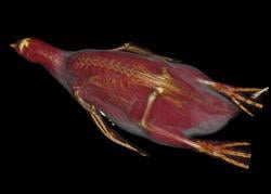
A 3-D CT scan reconstruction of a Humboldt penguin at the Brookfield Zoo in Chicago.
February 9, 2012 – A 3-D view does more than make for exciting movies in a theater; it can also improve the care of animals. The Chicago Zoological Society’s (CZS) Brookfield Zoo is the first North American zoo to use revolutionary 3-D imaging technology with on-site digital radiology and computed tomography (CT) equipment in its state-of-the-art animal hospital. The new technology allows the Society’s veterinarians to enhance two-dimensional CT scans, MRIs and ultrasounds with 3-D models that will enable them to better treat zoo animals.
The imaging will be particularly useful in the planning of surgeries, especially in difficult cases that were impossible to treat with 2-D imaging. Zoo veterinarians have already aided several animals in ways never before possible. For Hoover, a 17-year-old aardvark, a 3-D scan revealed that a hole from a missing tooth was draining into a sinus cavity. The discovery would have been impossible without the new imaging, and veterinarians are now monitoring the condition to prevent future health complications
Another animal benefiting from the technology is Pilgrim, a 9-year-old African crested porcupine, who requires regular tooth trims. Animal care specialists were at one point questioning whether one of Pilgrim’s incisors, which continued to overgrow, should be extracted. The 3-D imaging was helpful in showing why this extraction would be potentially risky and more difficult than the continued trimming. The 3-D renderings showed the anatomy of the incisors and their very close proximity to many other structures that would add to the difficulty of the procedure.
“This is exciting new technology that gives us much more information about our animals here than we’ve ever had before. We can use this information to improve our treatment and for proactive care to help ensure their well-being,” said Tom Meehan, DVM, vice president of veterinary services for CZS. “The more we understand — and see an animal’s anatomy — the more we enhance our ability to provide the highest quality of care.”
Meehan added that the technology provides a better visualization of various tissue densities simultaneously, while CT and MRI scans allow the viewer to choose only one or the other. Veterinarians are already also using the imaging to better care for a California sea lion, a bald eagle, an addax and a Humboldt penguin.
Vizua, the company providing this imaging solution, first showcased the technology this past November at RSNA 2011 in Chicago. The Seattle, Wash.-based company provides affordable real-time imaging technology that allows technicians to convert previous CT scans into a 3-D form without needing to purchase new equipment or software. These 3-D interactive images can then be explored and shared instantly with doctors and their patients anywhere in the world just using their existing Web browser.
“We are very excited that Brookfield Zoo is the first animal hospital to utilize this technology, and we are working closely together to ensure excellent care for the animals,” added Jean-Manuel Nothias, director of Vizua Biomedical Partner Relations.
Since first using the technology, Chicago Zoological Society veterinarians have made several JPEG photos and movie files to illustrate their findings and track animal health on an ongoing basis.
These images will be valuable teaching tools for students and residents within programs at the zoo, including the Society’s formal veterinary student training program through the University of Illinois College of Veterinary Medicine.
In addition to the new 3-D technology, the Animal Hospital at Brookfield Zoo also is adding new ultrasound equipment with support from the Aurelio Caccomo Family Foundation, which is committed to zoo animal well-being and care. The equipment will allow veterinarians to view images in greater detail and identify problems with smaller structures, which will be especially helpful when treating or diagnosing smaller zoo animals. The increased processing power will also capture moving images, such as images of the heart or circulation, in detail for the first time.
To view a video fly-through of the animals' 3-D reconstructions, go to www.itnonline.com/view-all/videos/?bclid=910141019001&bctid=1450625476001
For more information: www.CZS.org


 February 02, 2026


