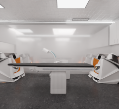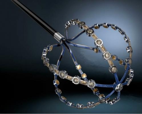
Image courtesy of Acutus Medical
October 6, 2015 — Acutus Medical, a global heart rhythm technology company, presented data that show the AcQMap High Resolution Imaging and Mapping System more rapidly and clearly maps atrial fibrillation (AF) than segmented computed tomography (sCT) mapping, the current standard for most AF ablation procedures. The initial study findings were presented during an oral presentation at the 10th annual Heart Rhythm Congress held in Birmingham, United Kingdom, Oct. 4-7, 2015.
The abstract titled, “Novel Global Ultrasound Imaging And Continuous Dipole Density Mapping: Initial Findings In AF Patients” was presented by Patrick Heck, M.D., Ph.D, Papworth Hospital, Cambridge, UK. The study evaluated the difference in mapping clarity and detection time between the AcQMap System and sCT in six patients diagnosed with complex arrhythmias. The findings concluded that the AcQMap System reconstructed an image of the atrium at 4.1 times higher resolution than that of sCT. The System also detected the arrhythmia more quickly than sCT (4:07 minutes versus 6:39 minutes, respectively).
“These data underscore the potential advantage the AcQMap System can provide to electrophysiologists (EPs) during AF procedures compared to current standards of mapping technology,” said Heck. “The image quality of the AcQMap System is unparalleled and allows EPs to see areas of the heart chamber we’ve never been able to visualize before. This is an impressive leap forward, truly opening the possibility to map AF with more precision to identify areas of interest as potential ablation targets.”
The AcQMap System can generate real-time ultrasound images of the heart chamber with quality equivalent to a CT or magnetic resonance image (MRI) in a matter of minutes. The system is the first and only to detect and display both the standard voltage-based maps as well as the higher resolution source charge (dipole density) maps through a proprietary algorithm. The result of combining highly accurate imaging with higher resolution mapping capabilities enables EPs to identify and locate the responsible charge for the arrhythmia, precisely target treatment areas and minimize unnecessary heart ablations.
For more information: www.acutusmedical.com


 January 29, 2026


