According to the Centers for Disease Control and Prevention, more than 70 million Americans currently live with ...
PET Imaging
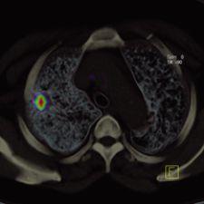
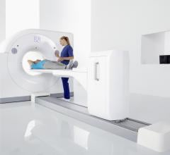
Positron emission tomography (PET) is a nuclear imaging technology (also referred to as molecular imaging) that enables visualization of metabolic processes in the body. The basics of PET imaging is that the technique detects pairs of gamma rays emitted indirectly by a positron-emitting radionuclide (also called radiopharmaceuticals, radionuclides or radiotracer). The tracer is injected into a vein on a biologically active molecule, usually a sugar that is used for cellular energy. PET systems have sensitive detector panels to capture gamma ray emissions from inside the body and use software to plot to triangulate the source of the emissions, creating 3-D computed tomography images of the tracer concentrations within the body.
August 1, 2007 - Positron Corp. announced that a recent study by The State University of New York at Buffalo ...
July 9, 2007 - A study published in the Journal of Nuclear Medicine reports that current PET-CT scanners with standard ...
Cardiac positron emission tomography (PET) is growing in popularity among cardiologists because it provides the ability ...
June 5, 2007 - GE Healthcare introduced at SNM a new version of the company’s Discovery Dimension designed to help ...

As in most partnerships, rarely are all things equal. Such is the case with hybrid PET/CT systems, where the vast ...
New SPECT/CT, cardiovascular IT and PET/CT technologies were on display by Philips Medical Systems at the American ...
GE Healthcare introduced its new 64-slice combination PET/CT system for cardiac imaging applications at the RSNA ...
Siemens has introduced a 40-slice configuration to its Biograph family of PET/CT systems. The Biograph family — which ...
A 64-slice combination PET/CT system for cardiac imaging, the Discovery VCT reportedly combines high-resolution ...
A revolutionary advancement in PET/CT, the GEMINI TF is the first PET/CT system that features time-of-flight PET ...
SceptreP3 is a hybrid PET/CT system that integrates the “Power of Three” suite of differentiating capabilities.
...


 September 09, 2007


