
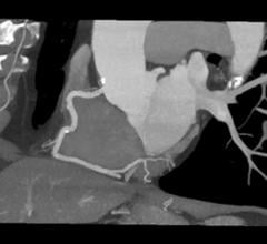
There were several interesting new trends in cardiovascular computed tomography (CT) imaging at the 2019 Society of ...
Cardiac Imaging
The cardiac imaging channel includes the modalities of computed tomography (CT), cardiac ultrasound (echocardiography), magnetic resonance imaging (MRI), nuclear imaging (PET and SPECT), and angiography.
August 6, 2019 — When used with a common heart scan, machine learning, a type of artificial intelligence (AI), does ...
 August 06, 2019
August 5, 2019 — A West Virginia-based rural medical outreach event showcased the use of point-of-care technology in an ...
August 05, 2019Sponsored Content

Feature
SPONSORED CONTENT — Studycast is a comprehensive imaging workflow system that allows healthcare professionals to work ...
June 17, 2025
Feature | FFR Technologies | Dave Fornell and Greg Freiherr
New technologies have been developed that may replace the traditional pressure wires and adenosine to assess the fractio ...
August 05, 2019
August 2, 2019 — The American Society of Radiologic Technologists (ASRT) announced its support for House Resolution (HR) ...
August 02, 2019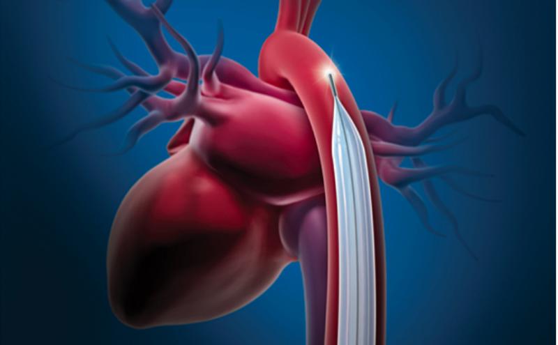
Feature | Dave Fornell, Editor
August 2, 2019 — Here is the list of the most popular content on the Diagnostic and Interventional Cardiology (DAIC) mag ...
August 02, 2019Sponsored Content
Feature | Cardiac Imaging
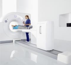
Cardiac positron emission tomography (PET) is growing in popularity among cardiologists because it provides the ability ...
March 05, 2024
News | Magnetic Resonance Imaging (MRI) | Jeff Zagoudis, Associate Editor
August 1, 2019 — The U.S. Food and Drug Administration (FDA) issued a new draft guidance titled Testing and Labeling ...
August 01, 2019
August 1, 2019 — Dassault Systèmes announced the five-year extension of its collaboration with the U.S. Food and Drug ...
August 01, 2019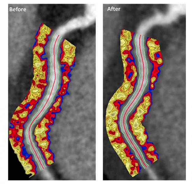
July 31, 2019 — Researchers found anti-inflammatory drug therapies used to treat moderate to severe psoriasis can ...
July 31, 2019Sponsored Content
Blog | Enterprise Imaging
As medical advancements continue to push the boundaries of what is possible in the field of structural heart ...
August 10, 2023
Nate Bachman, graduate research assistant in the Human Cardiovascular Physiology Lab of the Dept. of Health and Exercise ...
July 30, 2019
Mark Ibrahim, M.D., FACC, assistant professor of medicine and radiology, associate program director, advanced cardiac ...
July 26, 2019
Andrew Choi, M.D., FACC, FSCCT, co-director, cardiac CT and MRI, assistant professor of medicine and radiology, George ...
July 26, 2019Sponsored Content

Feature | Cardiac Imaging
Discover the key features of cardiovascular structured reporting that drive adoption, including automated data flow, EHR ...
November 07, 2022
Intelligent software solutions (aka deep learning, artifical intelligence, AI, machine learning), this seems to be ...
July 26, 2019
Pierre Qian, MBBS, cardiac electrophysiologist fellow, Brigham and Women's Hospital, explains how his facility is ...
July 25, 2019
Joao Cavalcante, M.D., FSCCT, director of structural heart CT and cardiac MRI, Minneapolis Heart Institute, discusses ...
July 24, 2019