March 31, 2021 — Heightened activity in the brain, caused by stressful events, is linked to the risk of developing a ...
Cardiac Imaging
The cardiac imaging channel includes the modalities of computed tomography (CT), cardiac ultrasound (echocardiography), magnetic resonance imaging (MRI), nuclear imaging (PET and SPECT), and angiography.
March 31, 2021 — ScImage Inc. celebrates its cloud partnership with Digirad Health after a year of successful deployment ...
March 31, 2021 — Driven by pandemic realities and clinical demand for portable and intelligent point-of-care ultrasound ...
SPONSORED CONTENT — Studycast is a comprehensive imaging workflow system that allows healthcare professionals to work ...

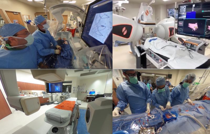
There is a lot of interest in how other hospitals organize and equip their cardiac cath labs and electrophysiology (EP) ...
March 23, 2021 — Researchers at Columbia University are using cardiac ultrasound to improve the critical need to ...
March 23, 2021 — Researchers at the Karolinska Institute in Stockholm, Sweden, have published the first-of-its-kind ...
Cardiac positron emission tomography (PET) is growing in popularity among cardiologists because it provides the ability ...
March 18, 2021 — GE Healthcare this week unveiled Vscan Air, a cutting-edge, wireless pocket-sized ultrasound that ...
March 17, 2021 — Around 50% of patients who have been hospitalized with severe COVID-19 (SARS-CoV-2) and have damage to ...

Heart failure (HF) is a prevalent yet silent epidemic, affecting 26 million people and costing global healthcare systems ...
As medical advancements continue to push the boundaries of what is possible in the field of structural heart ...

Intra-cardiac Echocardiography (ICE) uses catheter-based cardiac ultrasound array to image anatomy and devices inside ...

The latest cardiology practice-changing scientific breakthrough, late-breaking study presentations have been announced ...
March 8, 2021 — University of Kansas Health System Cardiologist Ashley Simmons, M.D., knew there could and should be a ...
Discover the key features of cardiovascular structured reporting that drive adoption, including automated data flow, EHR ...
March 3, 2021 — Impulse Dynamics recently announced that DEKRA Certification B.V., a global regulatory organization ...
February 25, 2020 — Results of a multi-centre, international, clinical trial co-led by Peter Munk Cardiac Centre (PMCC) ...
February 23, 2021 — A recent article in the Journal of the American Medical Association (JAMA) Cardiology demonstrated ...


 March 31, 2021


