A year after COVID-19 turned the world upside down, the American Society of Nuclear Cardiology (ASNC) asked members how ...
Cardiac Imaging
The cardiac imaging channel includes the modalities of computed tomography (CT), cardiac ultrasound (echocardiography), magnetic resonance imaging (MRI), nuclear imaging (PET and SPECT), and angiography.

May 27, 2021 — Philips Healthcare released a workhorse computed tomography (CT) system, the Spectral CT 7500, which has ...
Tom Jones, M.D., director, cardiac catheterization laboratories, Seattle Children’s Hospital, explains some of the new ...
SPONSORED CONTENT — Studycast is a comprehensive imaging workflow system that allows healthcare professionals to work ...

May 18, 2021 — Artificial intelligence (AI) derived heart measurements were able to predict COVID-19 (SARS-CoV-2) mortal ...
May 13, 2021 — The Centers for Disease Control and Prevention (CDC) just released a new statement relaxing the ...
May 12, 2021 — According to the British Heart Foundation, heart and circulatory diseases cause more than a quarter (27 ...
Cardiac positron emission tomography (PET) is growing in popularity among cardiologists because it provides the ability ...
May 12, 2021 — HeartFlow, Inc., a leader in revolutionizing precision heartcare, today announced that the National ...
May 11, 2021 — The American Institute of Ultrasound in Medicine (AIUM) and the American Society of Echocardiography (ASE ...
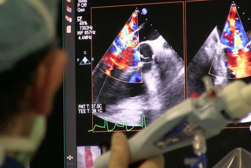
The resounding success of transcatheter aortic valve replacement (TAVR) has led the creation of hundreds of structural ...
As medical advancements continue to push the boundaries of what is possible in the field of structural heart ...
As a medical technology journalist, a little more than a decade ago I found myself sitting in those late afternoon ...
April 7, 2021 — Philips Healthcare announced U.S. Food and Drug Administration (FDA) 510(k) clearance for its Philips ...
Here are two quick clinical examples of point-of-care ultrasound (POCUS) lung imaging and cardiac imaging using a GE ...
Discover the key features of cardiovascular structured reporting that drive adoption, including automated data flow, EHR ...
April 1, 2021 – The ability to measure myocardial blood flow (MBF) as part of myocardial perfusion imaging (MPI) is one ...
Washington Health System (WHS) provides healthcare services at more than 40 offsite locations across three counties in ...
March 31, 2021 — Heightened activity in the brain, caused by stressful events, is linked to the risk of developing a ...


 June 02, 2021
