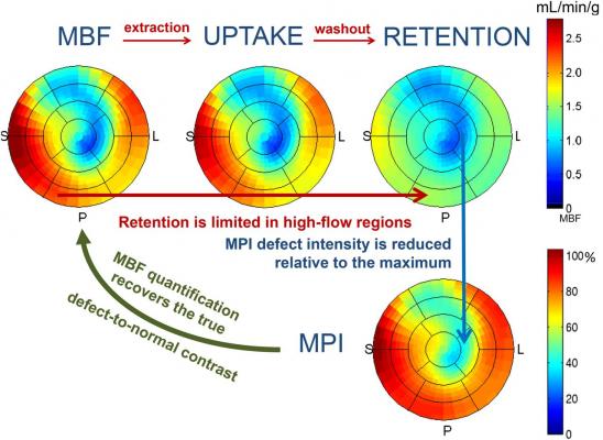
Polar maps demonstrating MBF, uptake, and retention along with their relationship to traditional relative MPI. Read more.
April 1, 2021 – The ability to measure myocardial blood flow (MBF) as part of myocardial perfusion imaging (MPI) is one of the most useful developments in contemporary nuclear cardiology. Now, a new practical primer provides clinicians with clear instruction for using this powerful diagnostic tool to improve patient care using positron emission tomography (PET) nuclear imaging. Developed by the American Society of Nuclear Cardiology (ASNC) and the Society of Nuclear Medicine and Molecular Imaging (SNMMI), “Practical Guide for Interpreting and Reporting Cardiac PET Measurements of Myocardial Blood Flow” was published online in the Journal of Nuclear Cardiology and the Journal of Nuclear Medicine.[1]
Measuring blood flow through coronary arteries provides clinicians with valuable information that helps them to determine the extent and severity of blockages that can cause a heart attack. MBF measurements also can identify microvascular disease, which occurs when the tiny arteries of the heart become blocked. In the past, microvascular disease has been difficult to diagnose using traditional imaging techniques. The information obtained through cardiac PET measurements of MBF also inform risk stratification, which helps guide treatment decisions.
“It is important that all cardiac PET providers be empowered to routinely incorporate quantification of myocardial blood flow as part of their daily workflow,” said Timothy Bateman, M.D., MASNC, a lead author of the writing committee composed of world-renowned leaders in cardiac PET. “While there are many excellent publications on all aspects of PET blood flow quantification, we felt that there existed a gap, especially for nuclear physicians practicing in non-academic centers. This practical guide was written to be a relatively easy read, and we hope will be helpful to expand implementation of blood flow measurements into daily clinical cardiac PET practice.”
The guide includes –
• An overview of the clinical value of quantifying MBF, including “six distinctive areas” of benefit;
• Tips for ensuring consistent high-quality MBF measurements;
• Essential technical requisites for accurate MBF quantification;
• Guidance on retention and compartment software models;
• A step-by-step approach to quality control for interpreting physicians;
• Recommendations for interpretation and reporting, including cutoffs for classification of risk and examples of useful sentences for reporting PET MBF;
• Explanation of challenges, such as for discordance between perfusion and MBF results;
• A glossary with key words and acronyms; and
• Case examples demonstrating how blood flow data should influence study results and clinical decisions.
Clinicians will find the guide to be both practical and user-friendly, with “important points” listed throughout the document, several figures and charts, and a dedicated section of case examples. Each case includes teaching points as well as recommended text to include to ensure reports will be helpful for referring clinicians.
“PET myocardial blood flow is perhaps the most important advance in nuclear cardiology in recent years,” said ASNC President Randall C. Thompson, M.D., FASNC. “The authors of this important statement for interpreting and reporting PET myocardial blood flow have succeeded in providing a clear and practical guide that lays out the essential issues for the practicing nuclear physician and technologist. This document is a valuable tool for learning the essentials of measuring PET myocardial blood flow, and it will be a helpful resource for practitioners at all levels.”
“Practical Guide for Interpreting and Reporting Cardiac PET Measurements of Myocardial Blood Flow” is available for download from the Journal of Nuclear Cardiology (JNC) and the Journal of Nuclear Medicine (JNM). For more guidance on using MBF, ASNC offers Cardiac PET Intensive Virtual Workshops, which includes three days of live case review, including a session dedicated to integrating MPI, MBF, and coronary calcium scoring.
About the American Society of Nuclear Cardiology: For over 25 years, the American Society of Nuclear Cardiology and its 4,000 members have been improving cardiovascular outcomes through image-guided patient management. As a #PatientFirst imaging society, ASNC establishes standards for excellence in cardiovascular imaging through the development of clinical guidelines, professional medical education, advocacy and research development. ASNC provides peer-reviewed original articles through its Journal of Nuclear Cardiology and operates the nation’s first noninvasive cardiac imaging registry, ImageGuide Registry®, to benchmark quality and improve patient care.
For more information: www.ASNC.org
Related PET Imaging Content:
VIDEO: Better Flow Quantification and Rise of PET Among Trends in Cardiac Nuclear Imaging — Interview with Rob Beanlands, M.D.
How to Start a Cardiac PET-CT Imaging Program
VIDEO: Practical Considerations for Starting a Cardiac PET-CT Program — Interview with Rupa Sanghani, M.D.
6 Trends in Cardiac Nuclear Imaging at the ASNC 2019 Meeting
Find more PET and other nuclear imaging news
Reference:


 January 05, 2023


