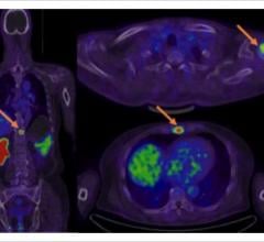
August 5, 2021 — A prospective, non-randomized, multicenter, first-in-human clinical study found good diagnostic and ...
Cardiac Imaging
The cardiac imaging channel includes the modalities of computed tomography (CT), cardiac ultrasound (echocardiography), magnetic resonance imaging (MRI), nuclear imaging (PET and SPECT), and angiography.
July 27, 2021 -- Cardiovascular diseases account for 32% of global deaths. Myocardial infarction, or heart attacks, play ...

Outside of medicine, computer-generated virtual twins of real machines like cars or airplanes have been used in ...
SPONSORED CONTENT — Studycast is a comprehensive imaging workflow system that allows healthcare professionals to work ...
July 26, 2021 – CathWorks announced that the Japanese Ministry of Health, Labour and Welfare (MHLW) has approved the ...
July 21, 2021 — Artificial intelligence (AI) medical imaging vendors Viz.AI and Avicenna.AI have partnered to enable ...
July 21, 2021 — Northwestern Medicine Bluhm Cardiovascular Institute recently became the first cardiovascular program in ...
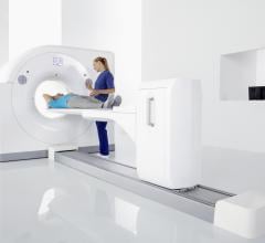
Cardiac positron emission tomography (PET) is growing in popularity among cardiologists because it provides the ability ...
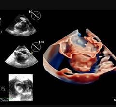
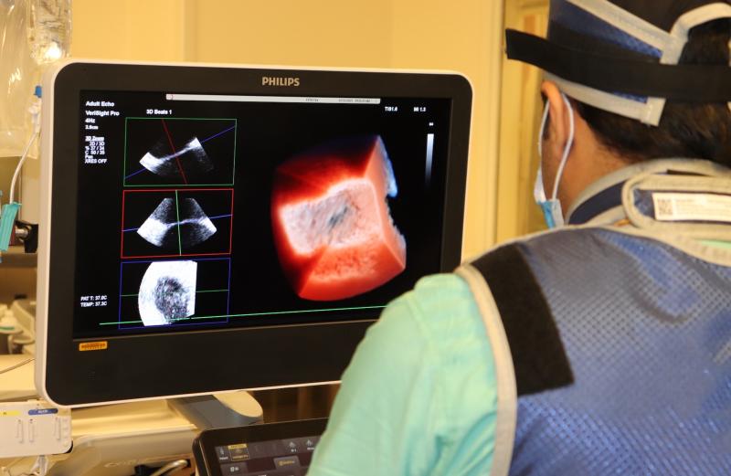
July 16, 2021 — Mayo Clinic recently became the first to use a next-generation 4-D intracardiac echo (ICE) imaging ...
July 16, 2021 — Canon Medical Systems USA is joining forces with Cleerly in a strategic partnership to support simple ...
July 15, 2021 — HeartFlow, which has commercialized noninvasive computed tomography derived fractional flow reserve (FFR ...
As medical advancements continue to push the boundaries of what is possible in the field of structural heart ...
July 14, 2021 — Performing the first cardiac scan on their new photon-counting detector computed tomography (CT) scanner ...
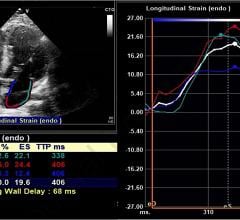
July 13, 2021 — Researchers at Johns Hopkins Medicine have shown that speckle-tracking strain echocardiograms may ...
July 13, 2021 — In a recent blog, the American Society of Nuclear Cardiology (ASNC) reported that Humana, one of the ...
Discover the key features of cardiovascular structured reporting that drive adoption, including automated data flow, EHR ...
July 2, 2021 — Philips has participated in an important research project to develop a magnetic resonance (MR) imaging te ...
Federico Asch, M.D., FASE, director of cardiovascular core labs, cardiovascular imaging, MedStar Health Research ...
June 29, 2021 – A new original science category being featured at the American Society of Echocardiography (ASE) 2021 ...


 August 05, 2021


