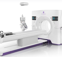
Toshiba's AquilionONE reduces contrast dose or coronary CTA.
The process of the coronary CT angiography (CTA) becoming the preferred imaging modality is akin to the process of obtaining a pilot’s license.
Before an individual gains the credentials to fly a Boeing 747, the aspiring pilot will be evaluated in myriad ways. Can the pilot take off in a cross wind? Can the pilot maintain control of the plane with a stalled engine? Has the pilot logged in the required amount of solo flight time?
Similarly, before CTA becomes universally recognized as the gold standard imaging modality for the coronaries, it must exhibit the necessary qualifications. Are both the positive and negative predictive values for coronary disease high enough? Has the radiation dose been reduced to an acceptable level? Can CTA be used for patients with renal insufficiency?
These are just some of the criteria that CTA must demonstrate before it “lands” the gold standard distinction. While many of these questions are still being debated, the recent innovations of CT systems and the development of appropriateness standards indicate that CTA will eventually overtake the invasive angiography for diagnostic imaging of the coronary arteries.
Evidence points to better performance
Technological advances in new CT systems are yielding better diagnostic results and opening up the scan to a more broad patient population, according to clinicians.
Joseph Schoepf, M.D., radiologist, professor, director of CT research and development at the Medical University of South Carolina in Charleston, has been working with cardiac CT since 1997. He primarily uses Siemens’ SOMATOM Definition CT system with dual-source technology for imaging the coronary arteries, a system that Dr. Schoepf believes has the best temporal resolution on the market.
“The most important improvement with the [SOMATOM Definition] was the temporal resolution,” Dr. Schoepf said. “The temporal resolution determines what sort of motion artifact you will get if you take pictures of a fast-moving organ such as the heart.”
Dr. Schoepf compares the temporal resolution on a CT scanner to the shutter speed on a camera. “If you have a camera with slow shutter speed and you try to take a picture of a fast-moving car, it’s very unlikely you’ll get crisp and clear images that are not blurred,” he said. “However, if you use a camera with a fast shutter speed, your image quality will significantly improve. The same is true for a CT scanner. It’s easy to understand why your shutter speed, (i.e. your temporal resolution), is the all-decisive factor that influences your diagnostic accuracy.”
According to Dr. Schoepf, the positive predictive value — the percentage of the time the scan found over 50 percent stenosis — at his institution has dropped from a 20 percent false positive rate on a four-slice CT to less than 5 percent with a 64-slice CT. Data measuring the SOMATOM Definition isn’t available yet, though he expects the system to yield good results.
Tony DeFrance, M.D., FACC, clinical associate professor at Stanford Medical School, director of cardiovascular imaging at the Nevada Imaging Centers, director of CVCTA Education and a member of the educational advisory board for Toshiba Medical Systems, is hopeful that the positive predictor value of Toshiba’s AquilionONE 320-slice system will improve when studies of that system are complete.
“My anecdotal experience from doing 200 cases is that it seems that we can see around the calcium quite a bit better with this machine with less blooming and artifact,“ Dr. DeFrance said. “I’m hopeful we’ll have less false positives.”
Dr. DeFrance said the AquilionONE allows clinicians to image patients with irregular heartbeats, a patient population that previous CT scans couldn’t always effectively image.
“[The AquilionONE] gets around that because it snaps the whole heart in a single heartbeat,” said Dr. DeFrance. “It doesn’t require six, or eight, or 10 heartbeats.”
Patient safety concerns
While high radiation exposure has been a concern among clinicians, a lengthy June New York Times article on CTA increased public awareness on the issue. Several clinicians feel the radiation risks associated with CTA are overstated, although they believe radiation levels must be minimized as much as possible without compromising diagnostic results.
Dr. DeFrance said patients must be exposed to less radiation before CTA can truly become the gold standard.
“We’ve made huge strides on this in the past six to 12 months,” Dr. DeFrance said. “We’re doing a lot more prospective imaging, so that’s just turning the tube on and off briefly, so you expose [patients] to about 80 percent less radiation.”
James Min, M.D., assistant professor of medicine, Weill Cornell Medical College at Cornell University, assistant attending physician, New York-Presbyterian Hospital in Ithaca, NY, who uses GE Healthcare’s new LightSpeed CT750 HD, has seen lower radiation on his system using the step and shoot method.
“Historically, I think the radiation for cardiac CT was about twice that of a normal chest CT,” Dr. Min said. “With the step and shoot method, it’s about 75 percent less than a regular chest CT.”
There also have been efforts to reduce contrast dose used in CTA. According to DeFrance, 64-slice scanners were able to get the rate down to a 70-80 cc dose. With the Aquilion One, the rate fell to a 40-50 cc contrast dose, said DeFrance.
“Once we’re getting down to the 40 or 50 cc threshold, we’ll be getting to a level that’s pretty acceptable for people with renal insufficiency,” Dr. DeFrance said. “It’s almost impossible with an invasive cath to get much lower than 40 or 50 cc dose.”
Redefining the cath lab
As CTA becomes more widespread, the cath lab will become more interventional in nature, according to Dr. DeFrance, who is also a board-certified interventional cardiologist. This is a positive change, Dr. DeFrance said, because fewer patients who ultimately don’t need an intervention will visit the cath lab, thereby decreasing complications associated with invasive catheterization.
According to Dr. DeFrance, recent studies have shown that CTA has cut diagnostic catheterizations by 10-15 percent, but increased the cath lab’s intervention volume by 20 percent.
“That’s going to become more important with current healthcare system,” he said. “They’re really going to hit cath labs hard that are doing low-acuity patients and make it not profitable with some of the Medicare and healthcare changes. Cardiac CT is going to be the gatekeeper for the cath lab.”
“As more data accumulates, CTA is going to become the gold standard for the workup of intermediate-and-low-probability patient chest pain, in my opinion, within two years,” he said.


 February 02, 2026


