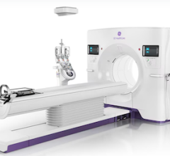
New computed tomography (CT) dose studies and growing public media attention have made minimizing unnecessary radiation dose to patients a priority for medical imaging facilities. In addition, state regulatory agencies and accrediting bodies are increasing their oversight and regulation of radiation dose. Reducing dose while maintaining good clinical image quality, however, is complex.
“Facilities are now being presented with a host of options from various vendors — dose tracking and analysis software, scanner hardware upgrades, technologist training, protocol optimization and more,” said W. Geoffrey West, Ph.D., president and chief medical physicist of West Physics Consulting, a national provider of medical and health physics consulting and testing services. “The radiation dose auditing and consultation services we have developed will: 1) assess the radiation doses a facility is starting with in each modality; 2) identify where improvements need to be made; 3) go ahead and make the most urgent and cost-effective improvements immediately with protocol optimization and enhanced staff training to reduce doses considerably; 4) quantify those improvements so the facility can inform patients, staff and referring physicians about their success; and 5) objectively evaluate how further available software and hardware upgrades would help lower doses even more.”
West Physics Consulting is one of many companies now offering CT dose reduction services. Another consulting provider is ECRI Institute, an independent nonprofit organization that researches the best approaches to improving patient care. It recently expanded its CT safety services to help hospitals and healthcare systems.
All CT manufacturers now offer versions of iterative reconstruction software to enhance the image quality of lower-dose CT scans. While differences in implementation exist, ECRI Institute clinical engineers determined through independent testing completed in 2011 that the overall effectiveness of the techniques is similar.
New Iterative Reconstruction Technology
Four of the major CT vendors introduced new iterative reconstruction software in 2012. GE Healthcare’s Veo iterative reconstruction technology can achieve clear chest CT images with less than 1 mSv of dose. Traditional chest CT scans can expose patients to anywhere from 5 to 10 mSv, and natural background radiation exposes the average American to around 3 mSv per year. GE also offers DoseWatch, a management tool that helps providers measure, track and optimize patient dose over time, and ASiR, GE’s standard iterative reconstruction software, available across GE Healthcare’s CT portfolio.
Siemens received U.S. Food and Drug Administration (FDA) clearance in June 2012 for the Somatom Perspective, an advanced 128-slice CT scanner. It uses the sinogram affirmed iterative reconstruction (SAFIRE) software to reduce dose by up to 60 percent compared to previous filtered back projection techniques.
Toshiba also released the newest version of its radiation dose reduction technology, Adaptive Iterative Dose Reduction 3D (AIDR 3D), in 2012, which is available on all new Aquilion CT systems.
Philips introduced the iDose4 Premium Package, which includes two technologies that can improve image quality — the iDose4 iterative reconstruction technique and metal artifact reduction for large orthopedic implants (O-MAR). Together, the technologies of the iDose4 Premium Package (iDose4 and O-MAR) produce high image quality with reduced artifacts.
Creating a Dose Index
While the CT scanner and reconstruction software technology is important, ECRI says dose monitoring and measuring also are critical to achieving lower radiation doses, explaining that one cannot control something that isn’t being measured. To facilitate dose monitoring, ECRI says a number of software options are becoming available, both from CT manufacturers and third parties. ECRI Institute believes these tools are just as important as CT scanner technology and will be vital for optimizing CT dose.
There are questions as to what are appropriate doses for various types of exams and patient sizes. In addition, not all facilities are on the same page with dose protocols or how they record dose. This prompted the American College of Radiology (ACR) to create its Dose Index Registry (DIR). In less than two years the registry has gathered data from more than 5 million CT scans that will help establish national benchmarks for CT dose indices.
The DIR is a radiology data registry that provides standardized, size-adjusted CT dose indices that facilitate meaningful comparisons, allowing imaging facilities to compare their CT dose indices to regional and national values. Information related to dose indices for all CT exams is collected, anonymized, transmitted to the ACR and stored in a database. Institutions are then provided with periodic feedback reports comparing their results by body part and exam type to aggregate results.
Once established, it is very likely the ACR dose indices will be incorporated into ACR appropriateness criteria software added to CT systems and PACS as part of clinical decision support/imaging ordering systems.


 February 02, 2026


