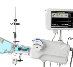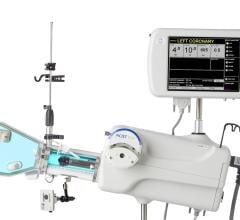
November 25, 2008 - MEDRAD is working on advancing the P3T software family of products for clinical applications beyond Cardiac CT, MEDRAD will demonstrate some of these potential advances at its RSNA Booth 2815 Hall A, South Building.
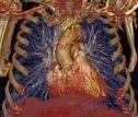
Personalized patient injection protocols are showing promise, according to a recent University of Pittsburgh Medical Center (UPMC) study which documented that contrast injection protocols personalized to patient-specific parameters of its test group resulted in a higher percentage of exams that could be classified as “diagnostic, without limitations” to rule out pulmonary embolism than resulted from non-personalized standard injection protocols. A diagnostic exam without limitations is the type that is most desired by radiologists. The personalized patient protocols provided better contrast enhancement of the pulmonary arteries in the study group as well.(2) Researchers presented the study results in a scientific poster at a meeting of the Society of Thoracic Radiology earlier this year. Copies of the study will be available at MEDRAD’s booth at the RSNA 2008 exhibition in Chicago, November 30 through December 5.
Joan Lacomis, radiologist at UPMC and co-author of the UPMC study, said, “Achieving a diagnostic CT image for pulmonary embolism, without limitations, helps the hospital and emergency department in a number of important ways -- with efficiency, patient care, and cost savings. Achieving high quality images for a higher percentage of studies multiplies these benefits and our research indicates that personalizing the contrast injection protocol to patient-specific parameters is a promising approach.”
Pulmonary Embolism (PE) is a sudden blood clot or blockage in a lung artery and is a serious condition. At least 100,000 cases of PE occur each year in the U.S., according to the National Institutes of Health. PE is the third most common cause of death in hospitalized patients, and if left untreated, kills about 30 percent of patients who have it. Most of those who die do so within the first few hours of the event. CT imaging is often used in the emergency department setting to quickly identify the presence of life-threatening disorders like PE, making efficiency important in quickly and accurately diagnosing the condition. CT has a high specificity and sensitivity in ruling out PE, and researchers in a 2005 meta-analysis (JAMA 2005; 293:2012-17) found that the negative predictive value of a CT angiography of the pulmonary arteries was 99.4 percent.
For more information: www.medrad.com and www.bayerhealthcare.com
(1) Lacomis, J; et al. “A Clinical Evaluation of the MEDRAD SmartFlow Power Injector Software System for Contrast Delivery in Patients Suspected of Pulmonary Embolism Undergoing Chest CTA”. Society of Thoracic Radiology. http://www.thoracicrad.org/meetings/str2008/Syllabus/additional/posters.pdf. March 2008.
(2) P3T Cardiac indicated for CT Pulmonary Angiography is pending FDA clearance [510(k) pending].


 January 11, 2024


