Vital Images Inc. featured its next generation of ViTALCardia, which includes a new EP planning tool and CathView, a new angiographic orientation.
Designed specifically for cardiologists, ViTALCardia is an advanced visualization and analysis tool designed for non-invasive evaluation of cardiovascular disease. The software includes several high-performance applications for the visualization and analysis of cardiovascular images, including: coronary vessel analysis, cardiac functional analysis, calcium scoring, peripheral vessel analysis and SurePlaque coronary plaque characterization software. With the availability of higher resolution image data, ViTALCardia focuses on the quantification and characterization of disease through the analysis of highly resolved image data.
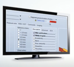
ViTALCardia’s CT Cardiac application now automatically probes, segments and labels the three main coronary arteries allowing for fast and easy non-invasive evaluation. With no user input, the application automatically segments the Left Anterior Descending artery, the Circumflex artery, and the Right Coronary artery. This anticipatory preprocessing facilitates a streamlined workflow. A new cardiovascular report was designed to automatically populate as findings are obtained, thereby reducing the number of steps needed to compile a final report.
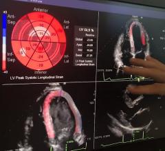
The powerful Cardiac Functional Analysis (CFA) application automatically calculates end diastolic and end systolic volumes to compute ejection fraction. Providing automatic qualitative and quantitative assessments of left ventricular function, CFA segments the left ventricle, automatically calculates cardiac output, myocardial mass, myocardial volume, and analyzes wall motion with 4D cine reviews. The application provides full color polar plots charting quantitative wall motion data.
ViTALCardia contains a 3-D advanced visualization and modeling tool for the electrophysiology (EP) lab. ViTAL EP, which is pending FDA clearance, automatically identifies anatomic landmarks, segments and probes the left atrium and the pulmonary veins to the first bifurcation. The application then creates a three dimensional anatomic model of the heart for super-imposing EP mapping. In addition to its powerful clinical features, ViTAL EP has a streamlined integration with the St. Jude Medical EnSite System.


 November 06, 2025


