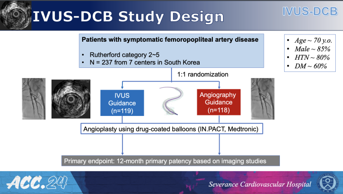
Results from the IVUS-DCB trial were recently presented at the American College of Cardiology Annual Scientific Session, ACC.24. Researchers found one-year success rates from angioplasty procedures, to open clogged arteries in the legs, were significantly higher among patients whose procedures were guided by intravascular ultrasound (IVUS) alongside angiography — compared with those whose procedures were guided by angiography alone.
April 11, 2024 — One-year success rates from angioplasty procedures to open clogged arteries in the legs were significantly higher among patients whose procedures were guided by intravascular ultrasound (IVUS) alongside angiography compared with those whose procedures were guided by angiography alone, in a study presented at the American College of Cardiology’s Annual Scientific Session.
The study, called IVUS-DCB, is the first randomized controlled trial to demonstrate the clinical benefits of using IVUS in angioplasty procedures for peripheral artery disease (PAD), a condition in which plaque builds up in arteries in the legs. IVUS is an imaging approach that enables visualization of the blood vessel wall and precise measurements of vessel size. The results suggest that the use of IVUS can help achieve longer-lasting benefits from the procedure.

Young-Guk Ko, MD
“IVUS is more accurate than angiography in measuring vessel dimensions. It facilitates achieving sufficient vessel lumen diameters and aids in evaluating the response of the target lesion to treatment,” said Young-Guk Ko, MD, cardiologist in the Division of Cardiology at Severance Hospital and Yonsei University in Seoul, South Korea, and the study’s lead author. “Despite the potential increase in procedural complexity, IVUS may benefit patients by enhancing treatment outcomes.”
The study focused on femoral artery disease, a form of PAD in which the femoropopliteal arteries that supply blood to the legs become narrowed or blocked, causing pain while walking. With angioplasty, operators inflate a small balloon inside the blood vessel, pushing the vessel open to restore proper blood flow. The balloons used in the study were also coated with drugs that help to prevent further plaque buildup.
To guide the placement of the balloon, most operators currently use angiography with a contrast agent to produce a 2D image of the vessel lumen. With IVUS, a tiny ultrasound device is inserted into the vessel via a catheter to produce cross-sectional and 3D images of the vessel for more accurate information on vessel dimensions and plaque morphology. Previous studies have shown IVUS can improve outcomes of endovascular procedures in the coronary arteries; the new study sought to assess whether the approach could bring similar benefits for procedures in the femoropopliteal arteries. Using IVUS alongside angiography entails additional costs because it involves additional procedural steps.
For the trial, researchers enrolled 237 patients undergoing angioplasty for femoropopliteal disease at seven sites in South Korea. Half were randomly assigned to receive IVUS plus angiography and half received angiography alone.
At 12 months, primary patency (a measure of the success rate of angioplasty and the study’s primary endpoint) was achieved in 83.8% of patients who received IVUS and 70.1% of those receiving angiography alone, a significant difference in favor of IVUS. Patients who received IVUS were also significantly less likely to require revascularization procedures and significantly more likely to show sustained clinical improvement compared with the angiography group.
Since IVUS is more accurate than angiography in measuring vessel dimension, researchers said the use of IVUS appears to have helped operators achieve a wider opening in the blood vessel for longer-lasting results.
“The IVUS group utilized larger pre-dilation balloon diameters and higher pressures prior to drug-coated balloon application as well as more frequent post-dilation and higher pressures for post-dilation after drug-coated balloon application, compared to the angiography group,” Ko said. “These optimizations, based on IVUS assessments, may have led to an increased final lumen diameter and better maintenance of the patent target vessel at the 12-month follow-up.”
One caveat is that most of the femoropopliteal artery lesions treated in the study were complex and extensive, stretching over 20 centimeters long on average, and it is unclear whether the benefits of IVUS would be the same for treating shorter and less severe lesions. In addition, the researchers said that the study only applies to procedures involving drug-coated balloons in femoropopliteal arteries, and further research would be needed to clarify possible benefits of IVUS with other types of devices, such as stents, and in other peripheral arteries.
The study was funded by Medtronic, Inc. and Korea United Pharm. Inc.
For more information: www.acc.org


 June 08, 2023

