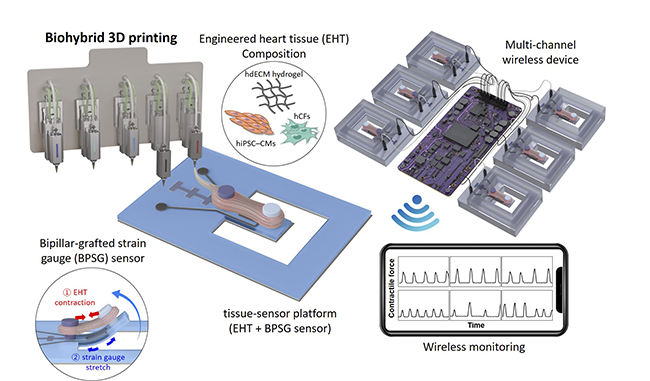
March 29, 2023 — Cardiotoxicity is a clinical condition that arises from using pharmaceutical agents such as antibiotics, which result in harmful effects on the heart, ultimately compromising its functional capacity. The left ventricle’s contractile ability is impaired, potentially leading to heart failure, a severe clinical outcome. Recently, a joint team of researchers from POSTECH and Georgia Institute of Technology successfully created an engineered heart via 3D printing technology that allows for early monitoring of drug-induced cardiotoxicity.
The team – led by Professor Jinah Jang (Department of Mechanical Engineering and the Department of Convergence IT Engineering), Uijung Yong (Department of Convergence IT Engineering), Donghwan Kim (School of Interdisciplinary Bioscience and Bioengineering), Professor Wan Kyun Chung and Professor Keehoon Kim (Department of Mechanical Engineering), and Professor Unyong Jeong (Department of Material Science and Engineering) at POSTECH along with a team from Georgia Tech led by Professor Woon-Hong Yeo and Dr. Hojoong Kim – has produced an engineered heart model using biohybrid 3D printing that enables in vitro monitoring of drug-induced cardiotoxicity in their research. The research findings detailing wireless, real-time, and continuous monitoring of drug-induced cardiotoxicity have been featured in the inside back cover of the latest issue of Advanced Materials, one of the most prestigious journals renowned for its coverage of material engineering.
Drug-induced cardiotoxicity is considered a major challenge in the early stages of drug development. While various in vitro platforms exist for preclinical cardiotoxicity testing, most conventional heart models lack physiological relevance and are less predictive of the cardiotoxic potential of drugs. Recently, 3D-engineered heart tissue (EHT) has been introduced as a promising alternative that emulates the heart’s physiological contraction and has been used by a number of researchers in their study of myocardial contraction and pharmacological effects. However, the absence of a suitable platform for continuous in vitro monitoring to test acute and chronic pharmacological effects remains an impediment.
The research team opted to use biohybrid 3D printing to engineer a heart model that surpasses traditional models. The team developed a platform integrating EHT with a bipillar-grafted strain gauge (BPSG) sensor. Their approach involved the 3D printing of two pillars that were subsequently grafted onto a substrate embedding strain gauges. After fabricated, the EHT was integrated into the BPSG sensor to create a tissue-sensor platform capable of real-time monitoring of heart contractions. Leveraging this platform with a multichannel wireless device, the team achieved continuous monitoring of EHT contractions and successfully demonstrated its utility in testing drug-induced acute and chronic cardiotoxicity.
Traditional in vitro monitoring systems for heart models’ contraction have had limitations in their ability to process large volumes of image-based data with high temporal resolution over prolonged periods. However, the tissue-sensor platform developed by the research team facilitates the quantitative measurement of contractile force with a relatively small volume of electrical readout, thus enabling long-term and continuous monitoring.
“This biohybrid 3D printing technique used to create the tissue-sensor platform has the potential in advancing drug development, representing a critical step towards the manufacturing of next-generation tissue-sensor platform,” remarked Professor Jinah Jang from POSTECH.
The study was conducted with the support from the Global Human Resources Project of the Ministry of Science and ICT and the Institute for Information and Communication Technology Promotion (IITP), the Regenerative Medicine Project of the Ministry of Science and ICT and the Ministry of Health and Welfare, the Alchemist Project for Breakthrough Technology Development of the Ministry of Commerce, Industry, and Energy and the Korea Evaluation Institute of Industrial Technology (KEIT), and the National Science Foundation (NSF).
For more information: https://international.postech.ac.kr/
Related 3-D Printing in Cardiology Content Content:
To Access the Online 3-D Printing and Printing Services Comparison Chart
VIDEO: Applications in Cardiology for 3-D Printing and Computer Aided Design
3-D Printed Heart Models Displayed at SCCT
Researchers 3-D Print Lifelike Heart Valve Models
Interventional Imagers: The Conductors of the Heart Team Orchestra
3D Systems Announces On Demand Anatomical Modeling Service
3-D Printed Models to Guide TAVR Improve Outcomes
Nemours Children's Health System Uses 3-D Printing to Deliver Personalized Care
Developments in Transcatheter Mitral Valve Replacement
1,000th 3-D Print Patient Treated at Henry Ford Health System


 February 03, 2026


