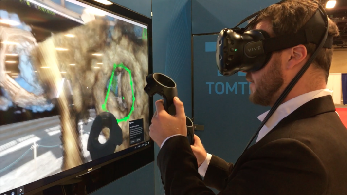
TomTec's work-in-progress virtual reality workstation to review echo exams in actual 3-D using video gaming technology. The operator is performing a valve area quantification measure during a demonstration on the expo floor of the American Society of Echocardiography (ASE) 2018 in June.
I attended the American Society of Echocardiography (ASE) 2018 meeting in June and have the following takeaways on cardiac ultrasound technology trends. To get an overview of the big picture trends I spoke with Sunil Mankad, M.D., FASE, the ASE 2018 meeting program chair and director of transesophageal echocardiography at Mayo Clinic, Rochester, Minn. He outlined these four areas:
• Advancement of 3-D echo;
• Point-of-care ultrasound;
• Integration of artificial intelligence (AI) and;
• New forms of image visualization
The first three areas have been major areas of discussion in sessions at ASE for the past couple years and are highlighted further below.
The last area of new image visualization includes the use of true 3-D imaging using special review stations with 3-D glasses, or use of virtual reality head visors. TomTec and GE Healthcare both showed work-in-progress virtual reality image review technologies in their booths at ASE to gather physician feedback on their potential utility.
The TomTec system allowed operators to select different image editing functions and to trace anatomy for quantification using handheld controllers. The operator works in a virtual environment where they can turn their head to find and physically grab images and tools. They use hand controller movements to rotate, zoom, crop and switch back and forth between 3-D and 2-D echo images of the same anatomy in the same view while reviewing an exam. The TomTec demonstration definitely won the title of the coolest new technology shown on the expo floor and was packed most of the meeting with people wanting to try it out.
Watch the VIDEO interview with Mankad, "4 Recent Advances in Echocardiography Technology."
Most Innovative New Echo Technology
I created a video tour of some of the most innovative new technology on the show floor of ASE 2018 meeting. The segments include virtual reality workstations, advanced 3-D cardiac ultrasound quantification and visualization, improved echo-fluoro image fusion technology and imaging aided by artificial intelligence. Watch the "Editor's Choice" VIDEO.
POCUS Becomes a Focus of ASE
A few years ago, ASE was concerned about the rapid expansion of point of care ultrasound (POCUS) for quick cardiac assessments by clinicians who are not trained sonographers or cardiologists. However, the society has changed that stance to fully embrace POCUS and is now looking to become the leader in this exploding area of imaging.
At ASE, I spoke with Michael Lanspa, M.D., director of critical care echocardiography services, Intermountain Medical Center, Salt Lake City, Utah, who is an advocate for POCUS training across medical departments. He serves on the educational committees for ASE, the American Thoracic Society (ATS) and the Society of Critical Care Medicine. He said there is a need for POCUS as a core educational mission, so ASE has decided to take this on. Lanspa said ASE sees this as a way to expand its expertise to new members looking for basic POCUS assessment training. Lanspa said echo labs already have the expertise and equipment on site to train staff from other hospital departments. He said this helps to add additional value for echo labs and help build interdisciplinary care teams within a hospital.
The society has run a hand-on POCUS seminar on the first day of its annual meeting for two years now, and includes it in several sessions throughout the meeting. The focus has been on basic cardiac assessments for non-cardiologists, lung assessments and basic ultrasound imaging training to speed triage of critical care patients, especially in the emergency room.
Watch the VIDEO interview with Lanspa "Point of Care Ultrasound (POCUS) in Cardiology and Critical Care."
3-D Echo Is Ready for Prime Time
The larger expense and lower frame rates of 3-D echo systems have limited their adoption over the past decade and have begged the question at previous ASE meetings whether 3-D is a necessary technology. However, sitting in on sessions at ASE 2018, the focus on 3-D imaging systems has definitely changed from a research tool to a front-line echo workhorse. Clinical evidence has shown 3-D echo allows for more precise measurements, better reproducibility between operators of varying skill levels, and the technology allows for automated quantification and an increasing role for artificial intelligence to greatly speed workflows.
Most 3-D systems are still operating below 30 frames per second, but the technology and speed is improving each year, said Lissa Sugeng, M.D., associate professor of medicine, director of echocardiography and director of the Yale Echo Core Lab, Yale School of Medicine. She believes the time has come where all echo labs need at least one 3-D echo system. She said these systems are needed minimally for cardio-oncology patient assessments. The imaging they provide is also valuable for surgeons and structural heart interventionalists who need the 3-D imaging for a more comprehensive assessment and visualization of valves, septal defects and the left atrial appendage.
As the frame rates improve and advantages in workflow are better appreciated, Sugeng said 3-D will likely begin to replace older 2-D systems in the coming years.
Watch the VIDEO interview "Can We Live in 3-D Echo?" with Sugeng at ASE 2018.
Artificial Intelligence in Cardiology
AI has become a key hot topic at all of the medical conferences I have attended over the past two years. AI products are now entering the market at a fast pace to augment the physician, mainly to improve workflow and complete complex analytical tasks that involve large amounts of data.
The top expert on AI at the ASE meeting was Partho Sengupta, M.D., DM, FACC, FASE, chief division of cardiology, director of cardiac imaging, West Virginia University Heart and Vascular Institute. He said AI is being integrated into echocardiography systems to speed workflow by identifying anatomy and automating quantification. However, he believes its larger impact may come from AI's ability to sift through large amounts of patient data from full echo studies to look for small variations from hundreds of exams to find new ways to diagnose disease prior to acute presentations. He said AI can also mine big data from hospital patient records to identify patients at high risk for various cardiovascular conditions long before they have symptoms.
Watch the VIDEO interview with Sengupta at ASE 2018.
Tricuspid Interventions Will be the Next Major Trend in Transcatheter Structural Heart Procedures
I spoke with echo guru Rebecca Hahn, M.D., professor of medicine and director of interventional echocardiography, Columbia University Medical Center, New York, at ASE about how imaging techniques, research on and development of new transcatheter therapies are all progressing hand-in-hand together. The "forgotten valve" is no longer being ignored, she said.
Device companies are rapidly developing transcatheter devices to treat tricuspid valve regurgitation. There has been a need to refine techniques for imaging the tricuspid valve to better understand disease progression and implantable device implantation and performance in the lower pressure side of the heart. She spoke on these topics during sessions at both ASE and at the Transcatheter Valve Therapies (TVT) conference the same weekend.
VIDEO: Should Student Athletes be Screened for Sudden Cardiac Arrest? — discussion with Malissa Wood, M.D.
360 View of EchoPixel's true 3-D Visualization Console at ASE 2018
Cardiovascular Ultrasound Technology Showcased at ASE 2018
360 View of a GE E95 Echocardiography System at ASE 2018
Recent Advances in Echocardiography Technology
360 Photo of Philips Epiq Cardiac Ultrasound System at ASE 2018

 June 12, 2024

