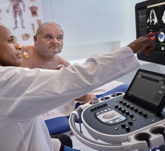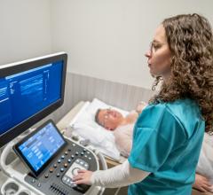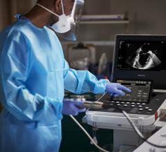
An example of one of the latest echocardiography image processing technologies, GE's Vmax.
Heart disease is globally pervasive and it is not going away. It’s the leading cause of death around the world, and the cause of one-third of all deaths in the United States.[1] That’s why the ability to detect cardiac complications early – with monitoring and medical imaging – represents a critical step in the future of improved patient outcomes.
A generation ago, cardiologists were looking at 2D, black and white images of their patients’ hearts — often missing critical diagnoses because of poor image quality. Since then, we’ve witnessed the emergence of 3D and 4D imaging, and software that can collect a practically infinite amount of data to create an image of the heart in a single beat.
Today, artificial intelligence (AI) presents a unique opportunity to make clinicians even more efficient.
By consuming, processing and analyzing massive volumes of data at extreme high speed, AI-powered solutions can support a variety of critical healthcare functions – from real-time assessment to point-of-care interventions and predictive analytics for clinical decision-making. For patients, these intelligent machines could mean shorter exam times and more confident diagnoses delivered more quickly to expedite quality patient care.
And while AI is often met with speculation in healthcare, AI-powered ultrasound systems with software-based architecture and GPU hardware engine allow cardiologists to read and review echo studies from a new perspective. AI allows links between observed anatomy, physiologic measurements and report interpretation are clearly visible and, for the first time ever, easily navigable.
MedStar Health’s Chief Scientific Officer Neil Weissman, M.D., FACC, FASE, noted, “As much as we have impacted the practice of medicine for the last 50 years, it's going to multiply over the next five to 10 years. With artificial intelligence, we're going to be more efficient and more effective at taking better care of the patients.”
For example, in a standard echo study, a sonographer will acquire hundreds of images. If a cardiologist wants to look at images from specific views of the heart, they need to review each individual clip – almost like flipping through a dense text book without a table of contents.
AI brings the potential for the cardiologist to indicate the specific structure so that the system may automatically pull all the images with that view, or identify specific anatomy and function of the heart bringing the exam review process in line with the clinician’s diagnostic questions – rather than managing a vast collection of images and measurements. This could save them critical time that they can now spend with their patients.
AI will enable cardiologists to spend more time focused on the patient’s condition to provide the best possible treatment options and outcome.
We are working towards a future world where heart disease is managed much earlier, more efficiently and will no longer take as many as 8.7 million lives in a single year. And with growing expertise and continued advances in technology, that vision may soon become a reality.
Editor's Note: Al Lojewski is the general manager of GE Healthcare's cardiovascular ultrasound division. This month, GE Healthcare is celebrating the 20th anniversary of the Vingmed acquisition, which introduced cardiovascular ultrasound to the GE.
Related ASE 2018 Technology Content:
Top Technology Trends in Echocardiography at ASE 2018
VIDEO: Editor’s Choice of the Most Innovative Echo Technology at ASE 2018
ASE videos, news and 360 images
Reference:


 June 12, 2024


