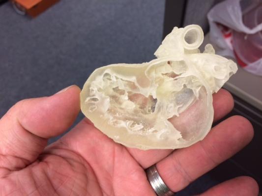
April 25, 2018 – Nemours Children’s Health System, a Florida-based health system with locations in six states, is now using in-house medical 3-D printing technology to create surgical models utilizing a U.S. Food and Drug Administration (FDA)-approved segmentation software. The software helps surgeons plan complex multidisciplinary cases in interventional radiology, cancer surgery and cardiac surgery. These models serve as a pre-planning blueprint and roadmap for Nemours surgeons and proceduralists, increasing confidence, reducing procedure times and minimizing unexpected findings while in the operating room.
“The simulation aspect of 3-D modeling is a game changer. To be able to look at a model of a tumor from all angles, without the restrictions of a 2-D image on a computer screen, is completely changing how we are planning complex surgery,” said Craig Johnson, DO, chair of medical imaging and enterprise director of interventional radiology.
Using modified models from volumetric 2-D computed tomography (CT) and magnetic resonance imaging (MRI), physicians can run simulations of the surgery and more accurately determine the tools they will need, with the multidisciplinary team involved, cutting down on waste. For example, certain models enable surgeons to drill holes in them to measure and select the appropriate medical equipment to use during the procedure.
“Three-dimensional modeling prepares us by helping us know exactly what we’re going to do. We do not have to plan on the spot if we come across something unexpected. Instead we’ve had imaging from radiology as well and the model,” said Tamarah J. Westmoreland, M.D., Ph.D., a pediatric surgeon at Nemours Children’s Health System. “The surgery is almost like a musical concert. It is rehearsed, planned and then executed without complication.”
The 3-D models are also invaluable in explaining procedures to patients and their families. An echo, MRI or CTR can be difficult for a parent to conceptualize. Instead, the Nemours team uses a true-to-size 3-D model of that patient. In many cases, the patients have the high-fidelity models next to them in their room as care teams explain their treatment plan.
“When we use a model to explain to a parent or a child a procedure, it’s clear, this approach is different,” Johnson explained. “They are able to visualize what we are going to do and it sets them at ease.”
Jessica Lewis is the mother of a Nemours patient and experienced this firsthand. Her 13-year-old son, Malachi, had a rare congenital coronary artery anomaly and needed cardiac surgery at Nemours.
“I was able to turn the model of my son’s artery around and look at it from all sides,” said Lewis. “The more educated you are about the procedure, the more empowered you feel because you completely understand what is going on with your child.”
The 3-D model printing service is yet to be reimbursable by insurance companies, but Daniel Podberesky, M.D., radiologist in chief at Nemours Children’s Health System, believes it is the right approach when it comes to providing the very best care.
“We are looking for ways to stay cutting-edge, all the while doing a better job clinically and helping families better understand what’s happening to their child,” said Podberesky.
For more information: www.nemours.org

 May 12, 2020


