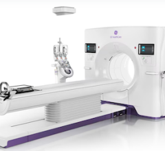
March 23, 2015 — Scientists from the U.S. Department of Energy’s Lawrence Berkeley National Laboratory have uncovered new clues about the risk of cancer from low-dose radiation. In this research, low-dose is defined as equivalent to 100 millisieverts or roughly the dose received from 10 full-body computed tomography (CT) scans.
They studied mice and found their risk of mammary cancer from low-dose radiation depends a great deal on their genetic makeup. They also learned key details about how genes and the cells immediately surrounding a tumor (also called the tumor microenvironment) affect cancer risk.
In mice that are susceptible to mammary cancer from low-dose radiation, the scientists identified more than a dozen regions in their genomes that contribute to an individual’s sensitivity to low-dose radiation. These genome-environment interactions only become significantly pronounced when the mouse is challenged by low-dose radiation.
The interactions also have a big impact at the cellular level. They change how the tumor microenvironment responds to cancer. Some of these changes can increase the risk of cancer development, the scientists found.
They report their research in the March 9 issue of the journal Scientific Reports.
Because mice and humans share many genes, the research could shed light on the effects of low-dose radiation on people. The current model for predicting cancer risk from ionizing radiation holds that risk is directly proportional to dose. But there’s a growing understanding that this linear relationship may not be appropriate at lower doses, since both beneficial and detrimental effects have been reported.
“Our research reinforces this view. We found that cancer susceptibility is related to the complex interplay between exposure to low-dose radiation, an individual mouse’s genes and their tumor microenvironment,” said Jian-Hua Mao of Berkeley Lab’s Life Sciences Division.
Mao led the research in close collaboration with fellow Life Science Division researchers Gary Karpen, Eleanor Blakely, Mina Bissell and Antoine Snijders, and Mary Helen Barcellos-Hoff of New York University School of Medicine.
The identification of these genetic risk factors could help scientists determine whether some people have a higher cancer risk after exposure to low-dose radiation. It could lead to genetic screening tests that identify people who may be better served by non-radiation therapies and imaging methods.
The scientists used a comprehensive systems biology approach to explore the relationship between genes, low-dose radiation and cancer. To start, Mao and colleagues developed a genetically diverse mouse population that mimics the diversity of people. They crossed a mouse strain that is highly resistant to cancer with a strain that is highly susceptible. This yielded 350 genetically unique mice. Some were resistant to cancer, some were susceptible and many were in between.
Next, Mao and colleagues developed a way to study how genes and the tumor microenvironment influence cancer development. They removed epithelial cells from the fourth mammary gland of each mouse, leaving behind the stromal tissue. Half of the mice were then exposed to a single, whole-body, low dose of radiation. They then implanted genetically identical epithelial cells, which were prone to cancer, into the fourth mammary glands that were previously cleared of their epithelial cells.
“We have genetically different mice, but we implanted the same epithelial cells into all of them,” said Mao. “This enabled us to study how genes and the tumor microenvironment—not the tumor itself—affect tumor growth.”
The scientists then tracked each mouse for 18 months. They monitored their tumor development, the function of their immune systems, and the production of cell-signaling proteins called cytokines. The researchers also removed cancerous tissue and examined it under a microscope.
They found that low-dose radiation didn’t change the risk of cancer in most mice. A small minority of mice was actually protected from cancer development by low-dose radiation. And a small minority became more susceptible.
In this latter group, they found 13 gene-environment interactions, also called “genetic loci,” which contribute to the tumor susceptibility when the mouse is exposed to low-dose radiation. How exactly these genetic loci affect the tumor microenvironment and cancer development is not yet entirely understood.
“In mice that were susceptible to cancer, we found that their genes strongly regulate the contribution of the tumor microenvironment to cancer development following exposure to low-dose radiation,” said Mao.
“If we can identify similar genetic loci in people, and if we could find biomarkers for these gene-environment interactions, then perhaps we could develop a simple blood test that identifies people who are at high risk of cancer from low-dose radiation,” said Mao.
The research was supported by the Department of Energy’s Office of Science.
For more information: www.lbl.gov


 February 02, 2026


