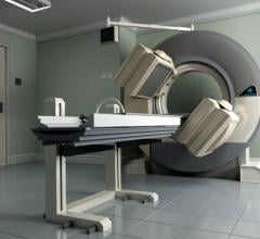
Single photon emission computed tomography (SPECT) remains a well-entrenched imaging modality for nuclear myocardial perfusion imaging (MPI) more than 30 years after its introduction. Due to SPECT’s reliability, cost-effectiveness and the wealth of data showing its clinical validation, it remains more common in MPI than its competition, positron emission tomography (PET). This is despite data showing PET’s much shorter scan times, fewer artifacts and better accuracy.
The high cost of the only FDA-approved, generator-based PET MPI agent, CardioGen-82, is a key factor keeping SPECT as the dominant modality. Cardiac PET took a further blow this past year when CardioGen-82 was abruptly pulled off the U.S. market in July 2011, after two patients set off radiation detectors at U.S. border crossings due to strontium breakthroughs. The agent uses a strontium (Sr)-82 generator to produce a rubidium (Rb)-82 radiotracer injection, but it requires close daily monitoring to prevent breakthroughs. The manufacturer, Bracco, is working with the FDA to bring the radiotracer back to market.
While SPECT technology has remained somewhat stagnant over the years, recent advances have improved the speed of exams and the diagnostic accuracy of the modality.
New gamma camera technology has allowed imaging times to be reduced from about 15-20 minutes down to about two to four minutes. Advances include both new camera design for better photon acquisition and sensitivity and better image reconstruction software.[1]
Wide beam reconstruction (WBR), a software program from UltraSPECT, reduces both dose and image acquisition time by 50 percent for SPECT MPI.[2] Researchers found that with either half the dose of Tc-99m sestamibi or half the acquisition time, WBR resulted in image quality superior to processing with today’s widely used OSEM (ordered subset expectation maximization) software. WBR’s reconstruction algorithm incorporates depth-dependent resolution recovery and image noise modeling to deliver a higher quality image with lower count density data.
Newer Systems
The adoption of combined SPECT/CT in one imaging system using a single patient table is growing. The combination of CT (computed tomography) offers anatomical imaging in addition to the functional molecular imaging of SPECT. In addition, CT is used for attenuation correction, resulting in better SPECT image quality and reduction in attenuation artifacts, especially in obese patients.
A major trend is the development of new dedicated cameras/detectors for cardiac-specific imaging. Spectrum Dynamics D-SPECT last year introduced a new-generation cadmium zinc telluride (CZT) detector that enables high-resolution, low-dose nuclear scans.
Frost & Sullivan recently recognized Siemens with the 2011 North America Product Differentiation Excellence of the Year Award for its IQ-SPECT system. It offers high-quality imaging by achieving maximum counts in one-fourth the amount of time required by conventional SPECT systems. The system features special collimators to focus on the heart, collecting up to four times more counts than conventional, parallel-hole collimators. A 3-D reconstruction algorithm models the position of each of the 48,000 collimator holes on each detector. This enables sophisticated distant-dependent isotropic (3-D) resolution recovery. It can complete a full SPECT scan in as little as four minutes.
GE Healthcare received U.S. clearance for the Brivo NM615 in January. The advanced single-head gamma camera enables lower amounts (up to 50 percent) of radiotracers and the potential for shorter exam times. This is made possible with a new detector design. The system is designed to handle larger patients with a 70 cm bore and table capable of handling patients up to 500 pounds. The system can be upgraded easily onsite to the Discovery NM630 or the Discovery NM/CT 670 SPECT/CT hybrid systems.
Comparison Chart
This story was an introduction to the SPECT and SPECT/CT comparison cahrt that appeared in the May-June 2012 issue of DAIC. To access the chart, click on the "Comparison Chart" tab at the top of the website page. Participants in the chart include:
Digirad - www.digirad.com
GE Healthcare - www.gehealthcare.com
Philips Healthcare - www.healthcare.philips.com
Siemens Medical Solutions - www.medical.siemens.com
References:
1. Tali Sharir, Piotr J. Slomka, Sean W. Hayes, et al. “Multicenter Trial of High-Speed Versus Conventional Single-Photon Emission Computed Tomography Imaging: Quantitative Results of Myocardial Perfusion and Left Ventricular Function.” JACC, May 4, 2010, Vol. 55, No. 18.
2. E. Gordon DePuey MD, Srinivas Bommireddipalli MD, John Clark CNMT, “A comparison of the image quality of full-time myocardial perfusion SPECT vs wide beam reconstruction half-time and half-dose SPECT.” Journal of Nuclear Cardiolog, April 2011, Vol. 18, No. 2.


 June 05, 2023


