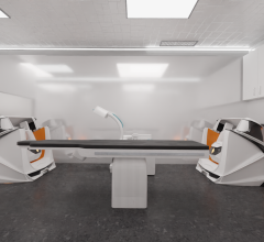
The Innova IGS series has two new additions.
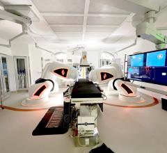
February 2, 2012 — GE Healthcare launched the Innova image guided system (IGS) 620 and 630, its next generation of electrophysiology (EP) biplane X-ray imaging. These systems offer high image quality, dose optimization with dedicated EP dose protocols, EPVision applications, and detector sizes optimized for 2-D and 3-D EP imaging.
Designed for a wide range of EP procedures, including complex ablations, Innova IGS 620 and 630 help provide the flexibility and reliability needed in an EP lab. These systems are part of a robust EP solution, designed to deliver a seamless flow of data throughout the patient visit. It serves as a platform for multi-modality image guided solutions, helping to guide diagnosis and treatment of complex heart disorders and arrhythmias.
Dose Management
Exposure to radiation during fluoroscopically guided procedures is a growing concern for both clinicians and patients. GE Healthcare offers three types of enablers to help address these concerns: core dose efficient technology designed to achieve imaging requirements while adhering to As Low As Reasonably Achievable (ALARA) principles, innovative dose reduction features, and dose management and reporting tools that can help optimize dose settings. New dedicated EP dose protocols on the Innova IGS 620 and 630 offerdefault settings optimized for EP procedures including low dose fluoroscopy.
EPVision Applications
Advanced 2-D and 3-D applications help electrophysiologists visualize anatomy and devices clearly and provide information to support case planning, real-time guidance and final assessment.
- Innova 3-D Rotational Angio enables physicians to quickly acquire and reconstruct a 3-D model of the left atrium for ease in navigation during procedures, while using dedicated EP dose protocols. These models can be easily exported to third-party 3-D EP mapping systems, helping to identify structures and differences in anatomy.
- Innova EPVision provides registration between 3-D models (3-D rotational angio, computed tomography [CT] or magnetic resonance [MR]) and 2-D fluoroscopy for 3-D anatomy visualization and device localization throughout procedures. Image stabilization features, including ECG gating and motion tracking to compensate for respiratory artifact, help provide a steady 3-D reference during complex procedures.
- The Innova IGS detector size is optimized for 2-D and 3-D EP imaging and real-time guidance. The panel sizes (dual 20 cm on IGS 620, 30 cm on IGS 630) can image the whole heart, and help facilitate rotational 3-D, taking advantage of a large field of view.
Workflow Enhancements
Innova IGS systems interface with the CardioLab recording system to streamline and integrate workflow and deliver advanced analysis tools to help enable clinical confidence. The Innova Central flexible, universal touchscreen panel lets the physician control certain CardioLab functions as well as Innova EPVision sequence storage from tableside for improved workflow.
GE’s 56-inch (diagonal) all-in-one large display monitor lets users see the information how, where and when they want it based on 120 predefined layouts. This single, configurable, high-resolution display is on a movable boom and offers 19 inputs to support many relevant data sources required during procedures. Users have tableside access to more than 120 pre-defined layouts and can customize a group of layouts according to procedure or their personal preferences by type of data, position on the screen, and image size (zoom up to 200 percent). Layout selection is fully integrated into the Innova Central Unit touch screen.
For more information: www.gehealthcare.com


 January 29, 2026


