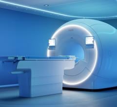
The Revo MRI SureScan Pacing System, approved by the U.S. Food and Drug Administration (FDA) earlier this year, was specifically engineered for MRI safety.
Estimates say up to 75 percent of patients with a pacemaker will need magnetic resonance imaging (MRI) over the course of their lifetime.[1] Yet as the industry has long been aware, MRIs can cause a number of adverse reactions when conducted on patients with a pacemaker. Aside from either the loss of pacing or inappropriate pacing, the MRI can cause current and heat to travel down to the heart muscle, resulting in local damage or life-threatening arrhythmias.
Traditionally, when faced with a pacemaker patient in need of an MRI, physicians and medical imaging professionals have turned to different diagnostic tests or have had to make a tough decision on whether the benefits of the MRI truly outweigh the risk.
Today, the market is changing, and patients have new device options that are significantly improving the relationship between pacemakers and medical imaging.
Tough Decisions
For healthcare professionals, the inability to perform an MRI on the many people in the United States with pacemakers can cause complications. Diagnoses or treatment decisions have to be made based on alternate imaging methods, and sometimes the risk of an MRI has to be taken.
When first presented with a pacemaker patient who needs an MRI, as professionals we ask ourselves what alternative testing can be done. Often it’s a computed tomography (CT) scan; the absence of magnetic fields has no adverse effect on the pacemaker or heart muscle. Yet with stroke, orthopedic patients or others with soft tissue conditions, an MRI is usually the best diagnostic test.
In these cases, we are faced with several options. The first, and most common, is to forego the MRI. An estimated 200,000 patients in the United States annually have to forego MRI scans because they have pacemakers.[2]
If it is determined the benefits of performing an MRI outweigh the risks, the patient and physician can choose to proceed with the MRI. In this scenario, both the pacemaker patient and the imaging professional acknowledge and accept the risk potential, and the MRI is performed with a cardiologist and crash cart present.
One other option to consider is removing the pacemaker system via surgical extraction with its associated risks. Yet understandably, many hospitals and imaging professionals do not wish to take on these risks.
MRIs can cause heat, static energy and radiofrequency energy, which can result in a number of adverse reactions, as previously mentioned. For the pacemaker, reactions include damage to the generator so that it no longer operates properly, or stops operating completely. And, for the patient, heat and current that travel to the heart muscle can cause significant damage.
Next-Generation Pacemaker Offers an Option
There is a new pacemaker system on the market that may completely change the traditional relationship between these devices and MRIs; in fact, the challenge of finding alternate diagnostic methods and the risk of conducting the MRI may be obsolete in a few years.
This new device – approved by the U.S. Food and Drug Administration (FDA) earlier this year – is the Revo MRI SureScan Pacing System. It was specifically engineered by the device maker for MRI safety and consists of the Medtronic Revo MRI SureScan pulse generator implanted with two CapSureFix MRI SureScan leads.
The system features several modifications on a traditional pacemaker to remove the risks we typically see with a traditional pacemaker and MRI. Medtronic shielded the most important components from heating by modifying the pacemaker generator and leads, adding additional shielding and running fewer but thicker electrode filaments in the leads.
As a result of these modifications, the Revo MRI SureScan contains several safety features, including SureScan Mode. When activated before an MRI scan, SureScan mode ensures patient safety in an MRI environment. The patient can be safely scanned, while the device continues to provide appropriate pacing.
Today, it is the only MR-conditional pacemaker approved by the FDA.
Firsthand Success
Recently, the MemorialCare Heart and Vascular Institute (MHVI) at Long Beach Memorial in Long Beach, Calif. – one of the state’s most comprehensive centers for the diagnosis, treatment and rehabilitation of heart disease – successfully implanted this new device into a male patient. Dealing with intermittent atrial fibrillation, slow heart rate and spinal cord irregularities, the care team determined it was imperative to first address the patient’s heart rhythm and implant a pacemaker.
The patient and care team chose the Revo MRI SureScan Pacing System, which allowed the most pressing arrhythmia to be dealt with, while also retaining the ability to examine spinal cord irregularities through an MRI in the future.
To date, the patient has not experienced complications with the surgery or device, and the MHVI has since implanted the device into a number of patients without complication. In fact, a clinical trial has been initiated since the release of the device to monitor its ongoing performance.
The Future of Imaging
While not yet widely used, this type of device is likely to become standard in a number of years. In terms of other imaging advances, we can expect to see defibrillators follow suit in the near future – the other main cardiac device that poses MRI risks. Yet with more complex circuitry and bigger components that allow it not only to pace, but also to charge energy and deliver a shock, modifying the defibrillator will be much more difficult than it was with a pacemaker.
At some point, however, it will be the standard-of-care to have MR-conditional devices implanted in all patients, whether a pacemaker or defibrillator. After all, in 2007, there were approximately 30 million MRI scans performed in the United States and its use continues to grow.[3]
As device makers continue to bring such options to market, MHVI will continue to lead the industry in being among the first to implant, use and provide feedback on such cutting-edge devices.
Editor's note: Serge Tobias, M.D., is medical director of electrophysiology at the MemorialCare Heart and Vascular Institute at Long Beach Memorial, Long Beach, Calif.
References:
1. Kalin R and Stanton MS. “Current clinical issues for MRI scanning of pacemaker and defibrillator patients.” PACE, 2005, 28:326-328.
2. Medtronic calculations cited in Rod Gimbel and Ted McKenna, “Safety of Implantable Pacemakers and Cardioverter Defibrillators in the Magnetic Resonance Imaging Environment,” Business Briefing: Long-Term Healthcare 2005 (2005) available at www.touchbriefings.com.
3. IMV, “Benchmark Report: MRI 2007,” IMV Medical Information Division.
Des Plaines, Ill., 2008.


 January 21, 2026


