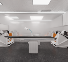
May 5, 2011 – During the Heart Rhythm Society’s 32nd Annual Scientific Sessions this week in San Francisco, GE Healthcare is showing a complete portfolio of integrated electrophysiology (EP) products featuring electrical signals recording and anatomical imaging through exceptional quality at the lowest dose, 3-D rotational angiography capabilities, and components integration. The GE solution provides the information electrophysiologists need to diagnose and treat difficult cardiac conditions and delivering it in a way that helps them focus on the patient, not the process.
The GE system allows a seamless flow of patient data from when the patient is admitted through treatment. The center of GE’s EP suite is the CardioLab Recording System. Built upon the Prucka legacy, it gives EP physicians the signal quality, data integration, and streamlined workflow they need—from the moment a patient is admitted, through diagnosis, treatment, and billing. Exceptional signal quality is key. CardioLab signal processing algorithms help provide exceptional waveform quality. And the high-performance CLab II Plus amplifier delivers the outstanding signal quality required for true bi-polar recordings with up to 224 inputs and 128 channels.
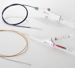
CardioICE combines EP recording with real-time intracardiac echo (ICE) images in one system, interfacing previously disconnected information to help physicians perform the most advanced EP procedures with confidence. Recording algorithms and workflow applications of CardioLab are combined with the compact, powerful GE Vivid i/q cardiovascular ultrasound system. Cardio ICE reproduces the Vivid i/q display in the CardioLab XTi window for continuous, real-time visualization of structures and devices synchronized with ECG waveforms. A remote control keyboard allows some Vivid i/q ultrasound parameters to be adjusted in the control room, removing the technician from the radiation field for increased operator safety.
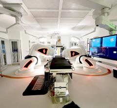
GE’s Innova is seamlessly integrated with CardioLab to facilitate streamlined workflow and deliver the advanced analysis tools that enable clinical confidence. The family of Innova X-ray systems delivers excellent clinical image quality and acquisition. And its dose efficient technology, dose reduction features and dose management tools help protect patients and physicians from radiation exposure. Independent studies confirm that when operating under live fluoroscopy, GE’s Innova doses are 22-75 percent potentially lower than other flat panel detector systems.[1]
Innova EPVision provides uncompromised registration between 3-D models (CT, 3-D angio and MR) and 2-D fluoroscopy for device localization throughout each procedure for device localization at all times. Image stabilization features such as electrocardiogram (ECG)-gated display or motion tracing in the image are offered for reduced image motion.
Innova 3-D Cardiac enables physicians to quickly acquire and reconstruct a 3-D model of the left atrium for easier navigation during procedures. These models can now be exported to all major 3-D mapping systems.
For more information: www.gehealthcare.com
Reference:
1. Patient Peak Skin Doses From Cardiac Interventional Procedures, Radiation Protection Dosimetry (2010) pp. 1–4, D. Zontar et al. “Radiation Dose of Interventional Radiology System Using a Flat-Panel Detector.” AJR (2009) pp. 1680–1685, K. Chida et al. “Comparison of the Radiation Dose in a Cardiac IVR X-Ray System, Radiation Protection Dosimetry.” (2010), pp. 1–7, Y. Inaba et al.


 January 29, 2026
