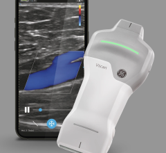
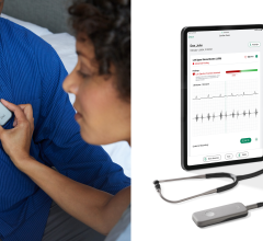
From the AHA exhibit hall this week, GE Healthcare has highlighted new breakthroughs to its Vivid cardiovascular ultrasound platform. The Vivid 7 Dimension ‘06 is designed to help clinicians assess cardiovascular anatomy and LV function with more accuracy.
GE spokespeople underscored the system’s ability to improve a clinician’s diagnostic confidence by making 4D cardiovascular imaging easier to use during day-to-day clinical exams.
“The additional information provided by 4D echocardiography increases diagnostic confidence for both the anatomical and functional assessment versus standard 2D imaging. The 4D protocol is both easy to use and easy to understand, and it is improving care across a broad range of patients,” said Dr. Randy Martin, professor of medicine and director of Echocardiology at Emory University Hospital in Atlanta, GA.
The Vivid 7 Dimension system is a PC based, software, raw data ultrasound platform that continues to evolve and improve year after year. Specific features comprised today include: Real-time 4D color flow full volume imaging, 4D LV Volume, Advance Tissue Synchronization Imaging (TSI, and Automated Function Imaging (AFI).
“AFI is a simple, user-friendly program that allows regional function quantification with minimal input,” said Dr. Ted Abraham, associate professor of Medicine and associate director of the Echocardiography Laboratory at Johns Hopkins University.
For more information, visit www.gehealthcare.com.


 January 28, 2026


