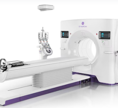
April 22, 2011 – The Society of Interventional Radiology (SIR) has a long-term commitment to radiation safety, taking a leading role in measuring and assessing radiation dosage; developing educational programs on radiation safety, radiation protection and reduction of skin dosage; and promoting the safety of patients and health care professionals. Four articles, published this month in the Journal of Vascular and Interventional Radiology, explore opportunities to improve patient safety through lower dosages and even new equipment that protects both patients and the diagnostic and interventional radiologist.
"The Society of Interventional Radiology views patient care and safety for patients and healthcare professionals among its absolute top priorities. To this end, SIR continually encourages clear communications between informed doctors and patients to recognize risks and benefits of radiation in diagnosis and treatment to save lives," stated SIR President Timothy P. Murphy, M.D., FSIR. "Interventional radiologists pioneered the procedures and standards for safely providing minimally invasive medicine. Patient safety is always incorporated into interventional radiology advances and treatments because interventional radiologists are highly trained in radiation safety, radiation physics, and the biologic effects of radiation and injury protection."
Murphy is an interventional radiologist and director of the Vascular Disease Research Center at Rhode Island Hospital in Providence. "The articles' unifying element is patient safety," said JVIR editor-in-chief Ziv J Haskal, M.D., FSIR. "By lowering a patient's exposure to damaging radiation—without compromising the treatment or diagnostic ability—and developing tools that provide improved functionality and protective properties for use by both the diagnostic and interventional radiologist, interventional radiologists again show why they are the driving force behind the development and implementation of the field's best practices,"
Haskal is also professor of radiology and surgery at the University of Maryland School of Medicine and vice chair of strategic development and chief of vascular and interventional radiology, image-guided therapy and interventional oncology at the University of Maryland Medical Center, both in Baltimore.
"Exploring ways to promote and practice safe radiation procedures and minimize radiation damage caused by medical imaging and image-guided treatments is of paramount concern to SIR members," said Murphy. The four new articles report on safety studies and examine new techniques in imaging used for diagnosis and treatment. The topics range from ways to optimize radiation dosages during fluoroscopic procedures, an examination of an ultra low-dose protocol for CT-guided lung biopsies, a comparison of a suspended protection system versus standard lead apron used by interventional and diagnostic radiologists, and an exploration of dosage differences during conventional CT guidance and a new cone-beam CT-guidance technique.
In "Optimizing Radiation Use During Fluoroscopic Procedures: Proceedings From a Multidisciplinary Consensus Panel," co-author James R. Duncan, M.D., Ph.D., Mallinckrodt Institute of Radiology, Washington University School of Medicine, St. Louis, Mo., noted that while fluoroscopic procedures have dramatically improved patient care and outcomes, the rapid rise in the use of ionizing radiation has renewed concerns about exposure during medical imaging. The SIR Foundation initiated a call for a multidisciplinary consensus panel on radiation use in 2010. The article spells out the panel's findings that in both diagnostic and interventional radiology the goal should be optimization during exposure—a strategy that recognizes the need to balance the risks versus the benefits of ionizing radiation.
The panel recommended the development of a registry to capture and analyze data from fluoroscopic procedures, recognizing the need for long-term support from both patient advocacy groups and federal agencies. Jason C. Smith, M.D., Loma Linda University Medical Center, Loma Linda, Calif., and his co-authors hypothesized in "Ultra-low-dose Protocol for CT-guided Lung Biopsies" that successful results could still be achieved during lung biopsies by radically lowering the CT dose. The study's conclusion proved that with an ultra-low-dose, or ULD protocol, the dose to the chest during CT-guided percutaneous lung biopsies is reduced greater than 95 percent from the standard protocol without decreasing technical success or compromising patient safety.
Daniel A. Marichal, M.D., Baylor University Medical Center, Dallas, Texas, and his colleagues evaluated the characteristics, reliability and ease of use of two radiation protection systems in "Comparison of a Suspended Radiation Protection System Versus Standard Lead Apron for Radiation Exposure of a Simulated Interventionalist." The group examined the effectiveness of a suspended radiation protection system that offers protection from the top of the head to the calves using a complex overhead motion system that eliminates weight on the operator and allows range of motion. When compared to the standard lead apron and tested on a mock interventionalist in a simulated clinical setting, the system greatly reduced exposures to many body areas.
A group of Dutch interventionalists, led by Sicco J. Braak, M.D., St. Antonius Hospital, Nieuwegein, the Netherlands, learned that using a recently developed needle intervention technique called cone-beam CT-guidance (CBCT) was effective and resulted in a considerably reduced dosage for patients when compared with traditional CT-guidance. Their results are presented in "Effective Dose During Needle Interventions: Cone-beam CT-guidance Compared With Conventional CT-guidance."
For more information: www.SIRweb.org


 February 02, 2026


