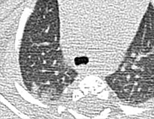
Chest CT findings of pediatric patients with COVID-19 on transaxial images. Male, 2 months old, 2 days after symptom onset. Patchy ground-glass opacities GGO in the right lower lobe. Image courtesy of Radiology: Cardiothoracic Imaging
May 5, 2020 — Children and teenagers with COVID-19 showed distinctive clinical and computed tomography (CT) findings, according to a new study published today in Radiology: Cardiothoracic Imaging. Compared to adults, pediatric patients generally had milder clinical symptoms, fewer positive CTs, and less extensive involvement on imaging. However, bronchial wall thickening was relatively more frequent on CT images from pediatric patients with COVID-19 in comparison with adults.
Among the study group, fever was less common in pediatric patients, and half the pediatric patients had only mild upper respiratory symptoms, such as cough or runny nose. Some had no symptoms at all.
A total of 61 patients, consisting of 47 adults (18 years old or older) and 14 pediatric patients (younger than 18 years old) with laboratory-confirmed COVID-19 by real-time reverse transcriptase polymerase chain reaction (RT-PCR) between January 25, 2020 and February 15, 2020 were enrolled in this study. All patients underwent chest CT within 3 days after the initial RT-PCR. The clinical presentation, serum markers, and CT findings were assessed and compared between the adult and pediatric patients.
Read the full study, Differences in Clinical and Imaging Presentation of Pediatric Patients with COVID-19 in Comparison with Adults.


 February 06, 2026


