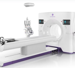
December 10, 2008 - New research has found that the availability of a portable eight-slice computed tomography (CT) scanner in an emergency room can significantly increase the number of stroke victims who receive a potentially life-saving treatment, according to results of a study conducted at North Shore Medical Center (NSMC)-Salem Hospital in Salem, MA, and presented at the annual meeting of the Radiological Society of North America (RSNA).
Siemens ARTISTE offers a comprehensive portfolio of image-guided and advanced treatment delivery tools, enabling clinicians to choose the appropriate treatment technique for each patient, make critical adjustments on the spot, and deliver true ART according to individual patient needs.
"The hospital's acquisition of a portable CT scanner facilitated more rapid assessment of acute stroke patients and is anticipated to increase the number of patients to whom thrombolytic therapy can be administered," said the study's lead author, David B. Weinreb, M.D., now a resident physician in the Department of Radiology at Hospital of Saint Raphael in New Haven, CT.
According to the National Institute of Neurological Disorders and Stroke (NINDS), stroke is the third leading cause of death in the U.S., and more than 700,000 cases of stroke are diagnosed annually. The most common kind of stroke, ischemic stroke, occurs when a blood clot blocks a blood vessel in the brain. Such strokes can be treated with thrombolytic therapy using a drug called tPA that dissolves the blockage. However, the window of opportunity to safely administer the medication is generally considered to be just three hours. Also, it is important to determine that there is no bleeding in the brain before administering tPA. One out of six strokes is caused by bleeding rather than clotting.
"tPA is usually the only shot we have at clot-induced ischemic strokes," Dr. Weinreb said. "But it needs to be administered in a closely monitored situation, because the drug can have extremely adverse effects in those patients whose strokes are instead due to bleeds."
Before a patient receives tPA, a head CT must be performed to ensure there is no bleeding in the brain. NINDS recommends that patients who arrive in the emergency room (ER) with signs of acute stroke undergo CT imaging within 25 minutes.
For the study, Dr. Weinreb and colleagues began using a portable CT scanner to assess stroke patients in the ER of NSMC-Salem Hospital. During the month prior to the acquisition of the portable scanner and for a four-month period following its installation, researchers measured how much time elapsed between a physician order for a head CT and performance of the scan.
The availability of the CT scanner in the hospital’s ER reduced the time between the order and exam from 34 minutes to 15 minutes, a reduction of 54 percent. Based on simulation modeling, the researchers estimated that this improvement would increase by 86 percent the number of stroke patients able to be treated with thrombolytic therapy within the three-hour window.
According to Dr. Weinreb, most stroke patients are taken to relatively small community hospitals where access to CT scanning may be limited. When a CT scanner is available, it is not always in proximity to the ER, making transportation of critically ill patients to the radiology department both difficult and time-consuming.
"A portable eight-slice CT can be easily added and used to accurately identify a head bleed in a stroke or trauma patient," Dr. Weinreb said. "This new technology is able to solve a very important problem for a community hospital, where the majority of stroke victims are being treated."
The co-author is James E. Stahl, M.D.
Disclosure: Dr. Weinreb consults as a medical writer for Neurologica.
Source: RSNA is an association of more than 42,000 radiologists, radiation oncologists, medical physicists and related scientists committed to excellence in patient care through education and research. The Society is based in Oak Brook, Ill. (RSNA.org)
For more information: www.radiologyinfo.org.


 February 02, 2026


