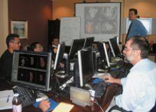
Students receive hands-on training with small classroom sizes at CVCTA Education.
Training physicians, especially cardiac specialists, has taken on an increasingly critical role. With more cardiologists, cardiovascular surgeons and vascular surgeons interpreting CT data, the need for proper training has increased exponentially in recent years.
CVCTA Education, based in San Francisco, CA, has embraced the need to train cardiac specialists and uses its expertise to expose doctors to a hands-on experience with the latest technologies utilized in CT angiography (CTA). Tony DeFrance, M.D., a board-certified interventional cardiologist and medical director of the training center, is at the helm of the training center’s comprehensive program — which he makes available at the center or on-site at a medical facility.
“CTA is an incredibly powerful procedure that impacts every level of cardiology,” said Dr. DeFrance. “Cardiologists are using sophisticated technology that makes the evaluation and treatment of coronary disease less invasive and less costly. Our mission is to educate physicians on how to efficiently perform, post-process and interpret CTA, including cardiac and peripheral vascular studies, using these technologies.”
Dr. DeFrance is also the director of Cardiovascular CT Angiography for Liberty Pacific Medical Imaging, a group of imaging centers with four locations throughout California. “Live” cases from these imaging centers are used in CVCTA’s courses.
Interpreting CTA is a completely new practice for many cardiac specialists; however, it is taking a vital role in the evaluation of coronary artery disease. Because it’s an emerging practice, the ratio of trained physicians to novice physicians is low, creating a significant demand for training. An essential component of learning to read cardiac CT studies is the need for hands-on workstation experience. It is not a skill that can be learned by watching DVDs or static images of someone else reviewing a study.
The training program offered at CVCTA is unique in that it is one of the only hands-on workstation programs in the country, and class sizes are kept small for personalized instruction and independent workstation experiences. It uses cutting-edge technologies including 64-slice scanners and two advanced visualization solutions, including Vitrea, offered by Minneapolis-based Vital Images. The four-day Part I course teaches students how to perform and interpret CT angiograms and familiarizes them with the “buttonology” of the workstations.
The Part II course exposes students to approximately 75 cases that vary in cardiovascular pathology. Level II certification is granted once the Part II course is completed and a total of 150 cases have been read. In addition, CME credits are given for each day completed, up to 32 credits for each level.
In addition, Dr. DeFrance recently rolled out the Part II CVCTA course in a self-paced software-based format through which the physician can learn the same material and techniques as in the class.
“It is difficult for busy physicians to be out of the office so we developed a software version of the level II training course,” said DeFrance. “A huge value to the Part I course is the hands-on component, but once students master the technology, they can easily complete the advanced course on their own schedule. We are thrilled to make essential education more accessible to cardiologists and radiologists.”


 January 09, 2026


