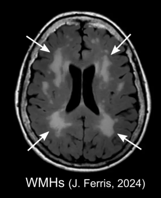
An example of white matter hyperintensities (WMHs) on a FLAIR MRI scan in an older adult. FLAIR MRI is a technique that is particularly good at detecting fluid in the brain. It helps researchers see areas of the brain that might be damaged by making the cerebrospinal fluid appear dark while the surrounding tissue remains light and detailed. This contrast makes it easier to identify abnormalities or injuries; WMHs appear as bright, patchy regions in the brain tissue. Image courtesy of Jennifer K. Ferris, PhD
May 7, 2024 — A new study by a global team of researchers, led by Sook-Lei Liew, PhD, of USC's Mark and Mary Stevens Neuroimaging and Informatics Institute (Stevens INI), has revealed that areas of age-related damage in the brain relate to motor outcomes after a stroke—a phenomenon that may be under-recognized in stroke research. The study was published online on May 3, 2024, in Neurology, the medical journal of the American Academy of Neurology.
A stroke often leads to motor impairment, which is traditionally linked to the extent of damage to the corticospinal tract (CST), a crucial brain pathway for motor control. Signaling along the CST is involved in a variety of movements, including walking, reaching, and fine finger movements like writing and typing. However, stroke recovery outcomes aren't fully predicted by damage to the CST, suggesting other factors are at play.
The new observational study from the Enhancing Neuroimaging Genetics through Meta-Analysis (ENIGMA) Stroke Recovery working group examines how one such factor could be white matter hyperintensities (WMHs)—areas of age-related damage in the brain's white matter, which represent vascular dysfunction and are known to impact cognitive functions. The goal of the ENIGMA Stroke Recovery working group, a National Institute of Neurological Disorders and Stroke (NINDS) -funded part of the ENIGMA Consortium, is to understand how changes in the brain after stroke relate to functional outcomes and recovery. ENIGMA Stroke Recovery has data from over 2,100 stroke patients collected across 65 research studies and 10 countries, comprising the most extensive multisite retrospective stroke data collaboration to date.
"We are grateful for our many collaborators around the world who lead independent stroke research programs and who are willing to come together and enable large-scale investigations into these critical questions about the role of overall brain health in stroke recovery and rehabilitation," says Dr. Liew, an associate professor at the Keck School of Medicine of USC, who also has joint appointments at the Chan Division of Occupational Science and Occupational Therapy, the Division of Biokinesiology and Physical Therapy and the USC Viterbi School of Engineering.
The study analyzed data from 223 stroke patients across four countries and found that larger WMH volumes were associated with more severe motor impairment after a stroke (e.g., difficulty moving or using their arm for daily tasks), independent of CST damage. WMHs are related to chronic hypertension, diabetes, high cholesterol, and smoking, among other factors and conditions, and have been strongly related to cognitive impairment, but not extensively studied in the context of motor impairment. Interestingly, the relationship between CST damage and motor impairment varied based on WMH severity. Patients with mild WMHs showed a typical relationship between CST damage and motor impairment, while patients with moderate to severe WMHs did not have this relationship. Instead, motor impairment was related to WMH volume, not CST damage.
These findings suggest that WMHs, indicative of cerebrovascular damage from a variety of sources, could provide additional context to understand an individual's potential for recovery post-stroke. Therefore, assessing WMH volume could improve predictive models for stroke recovery.
"WMHs are related to overall cardiovascular and brain health as we age. By integrating assessments of age-related brain health, we may be better able to predict stroke recovery and tailor rehabilitation to individual needs. This personalized approach could open avenues to improve outcomes after stroke," says Jennifer K. Ferris, PhD, of Simon Fraser University and the BC Centre for Disease Control, and lead author of the study.
The researchers' next step is to pursue longitudinal studies to confirm their findings. This insight lays the groundwork for developing more accurate markers for recovery, which could transform post-stroke care and rehabilitation.
"Dr. Liew and her team's work is integral to our mission to advance stroke research and discovery by using novel imaging technologies to study the structural and functional changes in the brain," says Stevens INI Director Arthur W. Toga, PhD. "We know that stroke is a leading cause of serious long-term disability, which has a considerable impact on public health. Studies such as this are a part of our goal to usher in a new era of precision rehabilitation that uses data-driven decision-making based on brain imaging and other noninvasive measures to identify personalized rehabilitation strategies.”
For more information: https://keck.usc.edu/

 January 28, 2026


