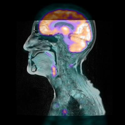
June 13, 2013 — Mirada Medical collaborators presentnew positron emission tomography/magnetic resonance imaging (PET/MRI) research.
PET/MRI is a rapidly developing field and Mirada is working to provide the software tools to support the research and trials necessary to realize the potential of the new modality in molecular imaging. The software supports both the hardware hybrid scanners and provides validated deformable registration for software based PET/CT(computed tomorgraphy)/MRI.
David Townsend, professor and director of the A*STAR-NUS Clinical Imaging Research Centre (CIRC) in Singapore, recently presented a paper entitled “Initial results of the assessment of PET/MR compared to PET/CT for diagnosis and staging of malignant disease.” This work is a comparative analysis of the image quality and quantification accuracy of hardware PET/MRI and PET/CT, a highly relevant topic of current interest in the field. Advanced quantification features and accurate deformable registration in Mirada PET/MRI software allowed the quantification of regions-of-interest simultaneously in both the PET/MRI and PET/CT of the same patient.
Another quantification analysis is presented in the work of Dr.Thomas Klausen of Rigshospitalet, Denmark, entitled “Image distortions and bias from dental restorations in PET/CT- and PET/MR-imaging.” In this work, Mirada’s support for multiple studies and multiple sequences enabled the analysis of artifacts arising from dental fillings on image quality and quantification on hardware PET/MRI and PET/CT.
Mirada’s software PET/MRI is an alternative to hybrid scanners and is based on deformable registration between the PET/CT and MRI to perform the fusion. A high-quality registration is critical for effective fusion between PET/CT and MRI. Mirada’s deformable registration algorithm, is the subject of another paper by Dr. Martin Lodge of Johns Hopkins University entitled “Combined PET/CT/MRI of the pelvis using software registration.” This work investigates the accuracy of the deformable registration between the CT and MR in the pelvis.
For more information: www.mirada-medical.com


 March 31, 2017

