Gregg W. Stone, MD
Mount Sinai Health System
March 31, 2025 — Using intravascular imaging (IVI) to guide stent implantation during complex stenting procedures is safer and more effective for patients with severely calcified coronary artery disease than conventional angiography, the more commonly used technique.
Those are the findings from the largest clinical trial of its kind comparing the two methods during percutaneous coronary intervention (PCI). The “ECLIPSE” trial results were presented at the American College of Cardiology Scientific Session (ACC.25) in Chicago. These results could shift treatment options for high-risk patients.
“The ECLIPSE trial shows that use of IVI to guide coronary stenting in severely calcified lesions prevents death, stent thrombosis, and unplanned repeat procedures in this high-risk patient population. These results extend the strong recommendations from recent U.S. and European societal guidelines that intravascular imaging with either optical coherence tomography (OCT) or intravascular ultrasound (IVUS) should be routinely used during complex coronary stent procedures,” says first author Gregg W. Stone, MD. Dr. Stone is Director of Academic Affairs for the Mount Sinai Health System and Professor of Medicine (Cardiology) and Professor of Population Health Science and Policy at the Icahn School of Medicine at Mount Sinai, and the study chair of the ECLIPSE trial. “Currently, IVI is performed in only 20 to 25 percent of these cases in the United States. I suspect its use will rapidly accelerate given study after study now showing reductions in death, stent thrombosis, and nearly every other adverse outcome after PCI when intravascular imaging is used.”
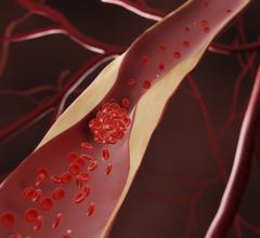
Patients with coronary artery disease—plaque buildup inside the arteries that leads to chest pain, shortness of breath, and heart attack—often undergo PCI, a non-surgical procedure in which interventional cardiologists use a catheter to place stents in the blocked coronary arteries to restore blood flow. About one-third of these cases in the United States—hundreds of thousands a year—have moderate or severely calcified lesions, where calcium builds up in the arteries. Ten percent of those cases are severe, meaning the blood vessels in the arteries essentially turn to bone, making the stenting procedure more challenging and higher-risk. Interventional cardiologists most commonly guide the PCI catheter by using angiography, which involves a special dye (contrast material) and X rays to see how blood flows through the heart arteries to highlight any blockages.
Angiography has limitations that make it difficult to determine the true artery size and the makeup of the plaque, and is suboptimal in determining whether the stent is fully expanded post-PCI and identifying other complications that affect the safety and effectiveness of the procedure. These limitations are amplified in calcified coronary arteries. By creating detailed two-dimensional cross-sectional images and three-dimensional views of the coronary arteries and blockages, IVUS and OCT provide a more accurate and specific picture of the coronary arteries than when the coronary angiogram is used alone. These high resolution imaging techniques can also better assess the adequacy of the implanted stent than angiography. Together, both imaging techniques allow for a more accurate measurement of vessel and plaque dimensions and composition and better assessment of stent implantation than can be read from the coronary angiogram.
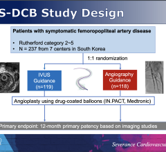
In the ECLIPSE study, researchers analyzed the outcomes of PCI in severely calcified lesions of 2,005 patients to see if IVI guidance improves survival without adverse cardiac events compared with angiography guidance. Patients with severely calcified coronary lesions were randomized at 104 sites across the United States. One group, 1,246 patients or 62 percent, had PCI with either OCT or IVUS guidance, and the other 759 patients, or 38 percent, had PCI with angiography guidance. The primary endpoint was the one-year rate of target vessel failure, the composite occurrence of either cardiac death, target-vessel myocardial infarction, or ischemia-driven target-vessel revascularization.
Overall rates of target vessel failure were 26 percent lower among patients who had IVI guidance than among those with angiography guidance. Researchers also observed a significant reduction in all-cause death, stent thrombosis, and target lesion and vessel revascularization among patients in whom intravascular imaging was used compared with angiography.
Researchers further analyzed the two imaging modalities — IVUS and OCT — to determine whether one was more effective. In the unadjusted analysis, patients had better outcomes with OCT compared to IVUS; however, the differences between the two were no longer significant when adjusting for factors including age, diabetes, and the number and severity of lesions.
“Overall, both OCT and IVUS were effective; additional studies are required to determine whether OCT is more beneficial in severely calcified lesions,” Dr. Stone adds. “Regardless, IVI with either OCT or IVUS should be used rather than angiography guidance to guide PCI in patients with severely calcified lesions.”
The ECLIPSE trial was funded by Abbott Vascular, Inc. (Abbott), Santa Clara, CA.
Source: Mount Sinai Health System

 July 31, 2024