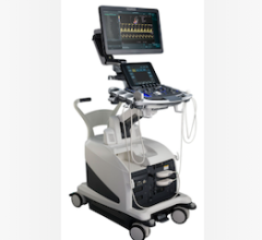
Cedars-Sinai investigators are using AI to analyze images from a common heart test to identify signs of valve disease. Photo: Getty.
April 16, 2025 — An artificial intelligence (AI) program trained to review images from a common medical test can detect early signs of tricuspid heart valve disease and may help doctors diagnose and treat patients sooner, according to research from the Smidt Heart Institute at Cedars-Sinai.
The work builds upon research published last year showing that an AI program can detect disease in the heart’s mitral valve by analyzing ultrasound images of the heart. For this new study, published in JAMA Cardiology, investigators applied AI to identify tricuspid regurgitation, a condition in which the heart’s tricuspid valve doesn’t close fully when the heart contracts, causing blood to flow backward, which can result in heart failure.

“This AI program can augment cardiologists’ evaluation of echocardiograms, images from a screening and diagnostic test that many patients with heart disease symptoms would already be getting,” said David Ouyang, MD, a research scientist in the Smidt Heart Institute, an investigator in the Division of Artificial Intelligence in Medicine and senior author of the study. “By applying AI to echocardiograms, we can help clinicians more easily detect the signs of heart valve disease so that patients get the care they need as soon as possible.”
Investigators trained a deep-learning program to flag patterns of tricuspid regurgitation in 47,312 echocardiograms done at Cedars-Sinai between 2011 and 2021.
The program detected tricuspid regurgitation in patients and categorized cases as mild, moderate or severe. They then tested the program on echocardiograms that the AI program never saw before from additional patients who underwent echocardiography at Cedars-Sinai in 2022 and patients from Stanford Healthcare. The program predicted severity of tricuspid regurgitation with similar accuracy as cardiologists who evaluated echocardiograms and when compared with results from MRI images.
“Future studies will focus on obtaining even more specific information about valve disease, such as the volume of blood flowing backward through a valve, and predicting outcomes if patients undergo treatment for heart valve disease,” said first author Amey Vrudhula, MD, a research fellow at Cedars-Sinai.

Investigators in the Smidt Heart Institute are applying AI to a variety of cardiac imaging tests.
“A major advantage of AI algorithms is that they never get fatigued and have the capacity to identify valve abnormalities from large populations of patients, taking personalized cardiology to a whole different level,” said Sumeet Chugh, MD, director of the Division of Artificial Intelligence in Medicine and the Pauline and Harold Price Chair in Cardiac Electrophysiology Research.
Other Cedars-Sinai authors involved in the study include Amey Vrudhula, MD; Milos Vukadinovic, BS; Alan C. Kwan, MD; Daniel Berman, MD; Robert Siegel, MD; Susan Cheng, MD, MMSc, MPH.
Other authors include Christiane Haeffele, MD, and David Liang, MD, PhD.
The work was supported by the Sarnoff Cardiovascular Research Award, research grants R00 HL157421 and R01HL173526 and support from AstraZeneca Alexion, as well as consulting from EchoIQ, Ultromics, Pfizer and InVision.


 October 09, 2025


