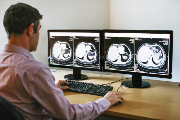
May 15, 2014 — Blackford Analysis is demonstrating its suite at the 2014 annual meeting of the Society for Imaging Informatics in Medicine (SIIM) in Long Beach, Calif. Blackford’s technology provides the ability to improve care and increase productivity by boosting image comparison efficiency by 20 to 50 percent.
Designed to be integrated directly into any image viewer, such as a PACS (picture archiving and communications system), advanced visualization viewer or universal viewer, Blackford Analysis’ products work seamlessly within existing systems to enable instant comparison of multiple image studies with a single click. By calculating and storing alignments with imaging studies, all healthcare professionals can benefit from instant study comparison.
Blackford Analysis software was used in the collection of data for a scientific presentation at SIIM 2014. The paper, entitled “PACS-integrated automatic deformable chest CT registration reduces radiologist time to match lung nodule locations across serial exams”, will be delivered by Matthew A. Barish, M.D., of Stony Brook Medicine, on May 16.
“Lung nodule studies can be quite a challenge to compare due to the differences in pulmonary anatomy caused by variance in patient positioning and breath hold,” said Barish. “Using a PACS integrated with Blackford Analysis has made a clear difference to our ability to quickly identify nodule locations across exams, and our early data confirms a significant reduction in the time spent locating nodules in follow-up studies allowing for quick determinations of nodule stability or growth.”
At SIIM 2014, Blackford Analysis is demonstrating four key elements of its software:
- Blackford MatchedCrosshairs enhances any image viewer to allow users to simply click once on a location in any scan to instantly find the same location in multiple scans from different timepoints and/or different modalities (computed tomography, magnetic resonance imaging or positron emission tomography).
- Blackford MatchedView enables any image viewer to automatically compensate for changes in patient positioning and acquisition planes between scans, automatically presenting views of compared exams in the same position and plane, and enabling like-for-like comparison.
- Blackford AutoSync gives image viewers the ability to perform slice synchronization across exams automatically, regardless of differences in acquisition protocol and patient positioning, so that reading can begin immediately when compared exams are displayed.
- Blackford Fusion enables image viewers to display accurate anatomical location of functional imaging findings by displaying fused views of exams from the same, hybrid or complementary modalities.
For more information: www.blackfordanalysis.com

 May 12, 2020


