Mark Ibrahim, M.D., FACC, assistant professor of medicine and radiology, associate program director, advanced cardiac ...
Computed Tomography (CT)
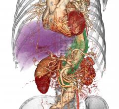
Cardiac computed tomography CT systems use a series of X-ray images to create an image volume dataset that can be sliced or manipulated on any plane using advanced visualization software. This channel includes content on CT scanners, CT contrast agents, CT angiography (CTA and CCTA), CT perfusion, spectral CT (also called dual souce or dual energy CT), and interative image reconstruction software that can reduce dose and make lower-quality CT images diagnostic.
Andrew Choi, M.D., FACC, FSCCT, co-director, cardiac CT and MRI, assistant professor of medicine and radiology, George ...
Pierre Qian, MBBS, cardiac electrophysiologist fellow, Brigham and Women's Hospital, explains how his facility is ...
As medical advancements continue to push the boundaries of what is possible in the field of structural heart ...
Joao Cavalcante, M.D., FSCCT, director of structural heart CT and cardiac MRI, Minneapolis Heart Institute, discusses ...
Arthur Agatston, M.D., clinical professor of medicine, Florida International University, Herbert Wertheim College of ...

One of the big trends in cardiac computed tomography (CT) imaging has been the introduction of noninvasive fractional ...
July 24, 2019 — The West Virginia University (WVU) Heart and Vascular Institute is the first hospital in the country to ...
Ron Blankstein, M.D., director of cardiac computed tomography, Brigham and Women's Hospital, and associate professor of ...
Quynh Truong, M.D., MPH, associate professor of radiology and medicine at Weill Cornell and director of cardiac CT ...
July 18, 2019 — Low doses of radiation equivalent to three computed tomography (CT) scans, which are considered safe ...
July 11, 2019 — Mednax Inc. and Mednax Radiology Solutions announced that Chief Medical Officer Ricardo C. Cury, M.D ...
July 10, 2019 — The Society of Cardiovascular Computed Tomography (SCCT) will present Stephan Achenbach, M.D., FSCCT ...
July 8, 2019 – Jonathon A. Leipsic, M.D., FSCCT, is the recipient of the 2019 DeHaan Award for Innovation in Cardiology ...
July 1, 2019 – vRad (Virtual Radiologic), a Mednax company recently made a scientific presentation, “Screening for ...
Roberto Lang, M.D., director of cardiac imaging at the University of Chicago, has been working with TomTec for the past ...


 July 26, 2019


