November 27, 2018 – Zebra Medical Vision and Clalit Health Services announced the completion of a research project that ...
Cardiac Imaging
The cardiac imaging channel includes the modalities of computed tomography (CT), cardiac ultrasound (echocardiography), magnetic resonance imaging (MRI), nuclear imaging (PET and SPECT), and angiography.
November 26, 2018 — Medical imaging software company Arterys will demonstrate its wide-ranging suite of artificial ...
November 25, 2018 — During the 104th Scientific Assembly and Annual Meeting of the Radiological Society of North America ...
SPONSORED CONTENT — Studycast is a comprehensive imaging workflow system that allows healthcare professionals to work ...
November 26, 2018 — HeartVista announced its artificial intelligence (AI)-driven, One-Click Autonomous MRI acquisition ...
Scott Schwartz, M.D., interventional radiologist and program director for IR residencies and the vascular and ...
November 19, 2018 – Medical artificial intelligence (AI) company Bay Labs and Northwestern Medicine announced that the ...
Cardiac positron emission tomography (PET) is growing in popularity among cardiologists because it provides the ability ...
DAIC Editor Dave Fornell takes a tour of some of the most innovative new cardiovascular technologies on display on the ...
November 15, 2018 — GE Healthcare announced it is recalling its Millennium Nuclear Medicine Systems due to an incident ...
Matthew Budoff, M.D., professor of medicine, David Geffen School of Medicine, UCLA, spoke at the 2018 American Heart ...
As medical advancements continue to push the boundaries of what is possible in the field of structural heart ...
November 6, 2018 — Siemens Healthineers announced the U.S. Food and Drug Administration (FDA) clearance of the Cios Spin ...
November 1, 2018 — Fujifilm SonoSite Inc. announced the launch of its redesigned SonoSite Institute, a comprehensive ...

November 1, 2018 — Here is the list of the most popular content on the Diagnostic and Interventional Cardiology (DAIC) ...
Discover the key features of cardiovascular structured reporting that drive adoption, including automated data flow, EHR ...
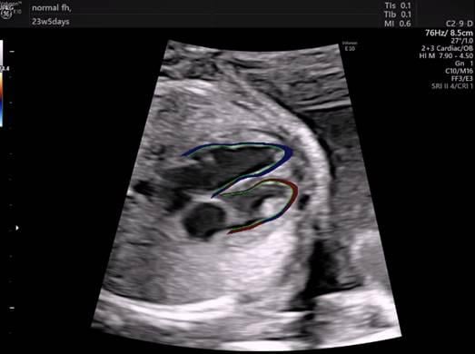
Example of GE Healthcare’s FetalHQ software for the ultrasound imaging of fetal hearts. The new tool runs on GE ...
October 31, 2018 — Vygon, an international group specialized in single-use medical devices, and French company ...

October 30, 2018 — At 18 weeks, a baby’s heart is the size of an olive and beating about 150 times per minute.[1] The ...


 November 27, 2018


