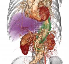
July 8, 2019 – Jonathon A. Leipsic, M.D., FSCCT, is the recipient of the 2019 DeHaan Award for Innovation in Cardiology ...
Cardiac Imaging
The cardiac imaging channel includes the modalities of computed tomography (CT), cardiac ultrasound (echocardiography), magnetic resonance imaging (MRI), nuclear imaging (PET and SPECT), and angiography.
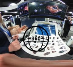
This 360 degree photo shows a basic, point-of-care cardiac echocardiogram being performed using a smartphone turned into ...
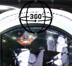
A view of a mitral valve on a GE Healthcare Vivid E95 cardiac ultrasound system scanned at the 2019 American Society of ...
SPONSORED CONTENT — Studycast is a comprehensive imaging workflow system that allows healthcare professionals to work ...
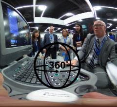
This is a 360 degree view of a live cardiac echo demonstration for the Siemens Healthineers Acuson SC2000 cardiovascular ...
This 360 degree photo is of a demonstration of AI capabilities being integrated into GE Healthcare ultrasound systems ...
July 2, 2019 — Philips recently announced new advanced automation capabilities on its Epiq CVx and Epiq CVxi cardiac ...
Cardiac positron emission tomography (PET) is growing in popularity among cardiologists because it provides the ability ...
Judy Hung, M.D., director of echocardiography, Division of Cardiology, Massachusetts General Hospital, Boston, discusses ...
July 2, 2019 — DiA Imaging Analysis has partnered with Konica Minolta Healthcare Americas Inc. to expand analysis ...
July 1, 2019 – vRad (Virtual Radiologic), a Mednax company recently made a scientific presentation, “Screening for ...
As medical advancements continue to push the boundaries of what is possible in the field of structural heart ...
Federico Asch, M.D., FASE, director of cardiac imaging research and director of the cardiovascular imaging lab, MedStar ...
Partho Sengupta, M.D., MBBS, chief of cardiology, West Virginia Heart and Vascular Institute, explains how wearable, sma ...
Sharon Mulvagh, M.D., FASE, FACC, FRCPC, professor of medicine, division of cardiology, Dalhousie University, Halifax ...
Discover the key features of cardiovascular structured reporting that drive adoption, including automated data flow, EHR ...
This is a quick example of how artificial intelligence (AI) is being integrated on the back end of cardiac ultrasound ...
Marielle Scherrer-Crosbie, M.D., Ph.D., director of echocardiography at the Hospital of the University of Pennsylvania ...
Federico Asch, M.D., FASE, director of cardiac imaging research and director of the cardiovascular imaging lab, MedStar ...


 July 08, 2019

