Arthur Agatston, M.D., clinical professor of medicine, Florida International University, Herbert Wertheim College of ...
Cardiac Imaging
The cardiac imaging channel includes the modalities of computed tomography (CT), cardiac ultrasound (echocardiography), magnetic resonance imaging (MRI), nuclear imaging (PET and SPECT), and angiography.

One of the big trends in cardiac computed tomography (CT) imaging has been the introduction of noninvasive fractional ...
July 24, 2019 — The West Virginia University (WVU) Heart and Vascular Institute is the first hospital in the country to ...
SPONSORED CONTENT — Studycast is a comprehensive imaging workflow system that allows healthcare professionals to work ...

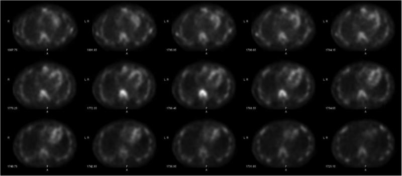
Cardiac amyloidosis is a highly morbid and underdiagnosed infiltrative cardiomyopathy that is characterized by the ...
Ron Blankstein, M.D., director of cardiac computed tomography, Brigham and Women's Hospital, and associate professor of ...
Quynh Truong, M.D., MPH, associate professor of radiology and medicine at Weill Cornell and director of cardiac CT ...
Cardiac positron emission tomography (PET) is growing in popularity among cardiologists because it provides the ability ...
July 18, 2019 — Low doses of radiation equivalent to three computed tomography (CT) scans, which are considered safe ...
July 18, 2019 — On June 30, 2019, the Centers for Medicare & Medicaid Services (CMS) announced the Johns Hopkins ...
July 16, 2019 – NorthStar Medical Radioisotopes LLC announced completion of construction on its 20,000-square-foot ...
As medical advancements continue to push the boundaries of what is possible in the field of structural heart ...
July 15, 2019 — The U.S. Food and Drug Administration (FDA) has approved Gadavist injection for use in cardiac magnetic ...
July 11, 2019 — Mednax Inc. and Mednax Radiology Solutions announced that Chief Medical Officer Ricardo C. Cury, M.D ...
July 10, 2019 — The Society of Cardiovascular Computed Tomography (SCCT) will present Stephan Achenbach, M.D., FSCCT ...
Discover the key features of cardiovascular structured reporting that drive adoption, including automated data flow, EHR ...

Here is an overview of the hot topics and new technology trends in cardiovascular ultrasound from the 2019 American ...
This is an example of a cardiac echocardiography exam performed using an iPhone and the Butterfly IQ ultrasound ...
This 360 degree photo shows a basic, point-of-care cardiac echocardiogram being performed using a smartphone turned into ...


 July 24, 2019


